|
|
Journal Home Contents Preview Next |
Pro Otology
Balkan Journal of Otology & Neuro-Otology, Vol. 3, No 1:46-48 © 2003
All rights reserved. Published by Pro Otology Association
Atresia of the External Auditory Meatus –
CT Findings in Two Cases
*R. Semova, *N. Traikova, †K. Dzhambazov
*Department of Roentgenology
†Department of ENT diseases, HMI, Plovdiv, Bulgaria
ABSTRACT
Objective: The aim of this study to present two cases with atresia of the external auditory meatus and the kneed of CT investigation in occasion of the forthcoming operation.
Study design: Report of two cases.
Setting: Department of Roentgenology and Department of ENT diseases, VMI – Plovdiv.
Patients: We present two cases in our study. The first case is of a 1-year-old girl with microtia of the right ear and data for soft tissue and bony atresia of the external auditory meatus. The second case is of a 15 years old girl with soft tissue atresia of the left external auditory meatus.
Interventions: In both cases CT investigations in the axial plain was made with following 2D and 3D reconstruction of the image. Then a surgical reconstruction of the ear canals was made.
Conclusions: The high-resolution computed tomography is the best roentgenological method for visualisation of atresia of the external auditory meatus when a surgical reconstruction of the ear is planed.
Key words: Atresion, External auditory meatus, CT investigation.
Pro Otology 1: 46-48, 2003
INTRODUCTION
The congenital anomalies of the ear include the anomalies of the external, middle and inner ear. They are bilateral in about 10 % of the cases. The embryonic development of the inner ear differs from that of the external and middle ear (1) wherefore the anomalies of the inner ear are not every time associated with the anomalies of the middle and external ear. Only the cases of severe chromosomal aberrations, which are caused from the teratogenic effects of any external factors, could make an exception from that. As the development of speech depends on the normal development of hearing the early determination of the congenital anomalies of the ear is very important, but is not easy every time (1). The clinical differentiation of the sensorial from the bony conduction could be difficult as well. The otoscopy is not possible when there is atresia or stenosis of the external auditory meatus. The success of the reconstructive surgery in that area depends to high extend on the early determination of the kind and degree of the anomaly. The main method for determination of the anomalies of the temporal bone and mostly the atresia of the external auditory meatus is the CT investigation (2, 3).
We present two cases with atresia of the external auditory meatus in which a CT investigation is made in occasion of the forthcoming operation.
MATERIAL AND METHODS
A CT “Sitek 1000” with standard and bony filter is used. The slices are 2 mm thin with a following 2D and 3D reconstruction of the image. The patients are scanned in an axial projection when lying on their back.
Case 1
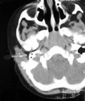
S. H. is one-year-old girl with microtia and clinical evidences for atresia of the right external auditory meatus.
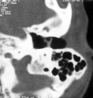
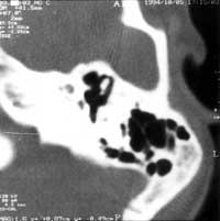
The CT investigation shows presence of 16-mm soft tissue atresia and a 6-mm bony atresia of the right external ear duct. The left ear canal is passable. The tympanic cavities are symmetrical with properly situated auditory chains on both sides. There are no pathological changes in the anatomical structures of the inner ear and mastoids (FIG. 1, 2, 3).
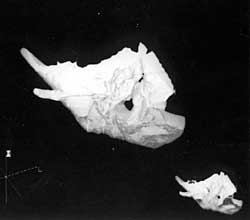
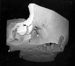
The area with the bony atresia is best visualized on the 3D reconstruction of the image (FIG. 4).
Case 2
D. T. is a 15 years old girl with microsomia and evidences for unilateral (left side) facial hypoplasia – hypoplasia of the maxilla, hypoplasia of the mandibular arm and atresia of the external auditory meatus.
No other bony anomalies were found from the roentgenological investigation.
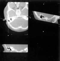
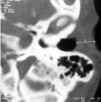
With the help of the CT we found only 10-mm soft tissue atresia of the left auditory meatus. There were no evidences for another anomalies of the temporal bone (FIG. 5A, 5B, 6, 7).
In both cases a surgical reconstruction was made and the normal structure of the canals was restored.
|
|
DISCUSSION
The atresia of the external auditory meatus could be bony or membranous. When it is bony no tympanic membrane could be found (6, 7). In such cases an atretic plaque forms the lateral side of mesotympanum. The deformation of the auditory ossicles is typical also the confluence between some of them and with the atretic plaque (mostly the malleus) (6). Some times there’s a missing part from the chain but it’s rare. The bony plaque could be widespread from tegmen to the auditory ossicles. The cavity of the middle ear is often constricted. Thеre are some roentgenological methods which are in use for diagnosing the atresia of the external auditory meatus – roentgenography of the area of pyramid and mastoid in the way of E. Mayer.
REFERENCES
Tomov A. Tomograph characteristics of changes in atresia of the external auditory meatus. Oto-rhino-laryngology 1982;5:28-9.
Fritz P, Rieden K, Lenarz T. Radiological evaluation of the temporal bone disease: high resolution computed tomtgraphy versus conventional x-ray diagnosis. The British Journal of Radiology 1989;62:107-13.
Jeakley J, Jahrsdoerfer R. CT evaluation of congenital aural atresia: What the radiologist and surgeon need to know. J of Computer Assisted Topmography 1996;20(5):724-31.
Schratter M, Swoboda H, Canigiani G, et al. Radiodiagnostik bei Schlafenbein-Anomalien. Radiologe 1988;28:481-8.
Von Koster O, Straehler-Pohl H-J, Kim K. Hochauflosende Computertomtgraphie bei Missbildungen des Gehor- und Gleichgewichtsorganes. Fortschr Roentgenstr 1987;147:39-45.
Eelkema E, Curtin H. Congenital anomalies of the temporal bone. Seminars in ultrasound, CT and MRI, Orlando 1989;10:195-212.
Mehra N, Dubey S, Mann Sh, et al. Correlation between high-resolution CT and surgical findings in congenital aural atresia. Arch Otolaryngol Head and Neck Surg 1988;114;137-41.
|
Pro Otology |
Journal Home Contents Preview Next |