|
|
Journal Home Contents Preview Next |
Pro Otology
Balkan Journal of Otology & Neuro-Otology, Vol. 4, No 1:2327 © 2004
All rights reserved. Published by Pro Otology Association
The Problems Encountered during Stapes Surgery
and Their Solutions
Nuri Ozgirgin
Faculty of Medicine, Bashkent University, Ankara, Turkey
ABSTRACT
Objective: Stapes surgery frequently has the potential of many complications related with the difficulties that can be only realized during the surgery. The solutions to the possible problems are discussed in this study.
Study design: Retrospective case review.
Setting: Bashkent University Hospital.
Patients: 90 patients with otosclerosis requiring operative intervention.
Interventions: 90 primary stapedectomies; 16 revision stapes operations.
Results: Among the 90 primary stapedectomies anatomic variations that have the potential of affecting the out come of surgery have been found in 15 cases. Prominent facial nerve was present in 4 cases, and the facial canal was dehiscent in 2 cases. The oval window was fully obliterated by the otosclerosis in 2 cases and it was narrow and deep in 7 cases. 16 revision stapes operations were performed and prosthesis dislocation and erosion of the long process of the incus were the frequent findings. Revision operations were also performed for two cases with reparative granuloma and one case with postoperative adhesive otitis media (cholesteatoma) as well.
Conclusions: The surgeon must be aware of the anatomical variations that may contribute to difficulties. These anatomical variations get importance when they especially involve the oval window, stapes footplate and the facial nerve portion that is close to the oval window. Other ossicular problems and the extent of the disease are other factors that closely affect the outcome of the surgery.
Key words: Stapes surgery, Anatomic variations, Intraoperative difficulties.
Pro Otology 1:2327, 2004
Introduction
Stapes surgery already carries the risks of additional hearing loss, tinnitus and facial nerve paralysis. Some difficulties that are met during the operation increase the morbidity of the surgery. Special attention should be given to the anatomical variations related to the stapes and the oval window during the operation. The deeply situated narrow oval window and facial nerve anatomic variations are the problems that are met frequently during the primary operations. Erosion of the long process of incus and dislocation of the prosthesis are the most frequent finding that requires late revision operations.
In this study the intraoperative difficulties that are met during the primary and revision stapedectomy operations are discussed with our suggestions to overcome the problem.
Material and Methods
90 primary stapedectomies as well as 16 revision cases were evaluated. Among the primary operations 15 cases showed intraoperative difficulties. Primary stapedectomies of six cases out of 16 revision operations belonged to the author.
The operative technique
During the operation, the surgeon has to be focused on the position of the oval window, the angle of the axis of stapes, the location and the situation of the facial nerve as these structures constitute the major anatomical factors that may affect the outcome of the surgery. Ossicular problems also have to be reminded.
The operation begins with Lemperts incision following local anesthesia. Following the elevation of the tympanomeatal flap, fine 45 degree needle is used to detach the annulus of the tympanic membrane from the bone. The stapedial tendon is excised either by using micro scissors or by argon laser with a power of one watt.
Crurectomy is performed through breaking them by using 45 degree needles. In case of using laser, the posterior crus is vaporized by applying 2 watts of power. It is generally difficult to manipulate the anterior crus too by using laser because of its position. However, the anterior crus is weaker and has lower resistance to mechanical stress so it is easily fractured following the removal of posterior crus. In case of performing the crurectomy by using the needles, there is always a risk of dislocation of the footplate.
The creation of the opening in the footplate is the most important stage of the operation. Fractured or floating footplate creates a great risk for hearing. This is likely to be occurred when using needles or hand held drills. If the footplate is fractured, then a hemi or total stapedectomy has to be planned. Lasers are superior to mechanical instrumentation as they do not apply any pressure on the footplate and the annular ligament. The diameter of the argon laser beam is about 150 microns, so if a rosette is formed by performing 3 to 5 applications (shoots) an opening that tightly hosts for 0.6 mm piston can be created. The tiny laser openings are easily united by using a needle. The char-coal is aspirated.
The argon laser is a powerful instrument on performing small fenestra stapedotomies. The concentrated heat energy created by the laser vaporizes the target tissue without any bleeding and this maintains the ideal conditions for the stapes footplate. It facilitates the surgery and shortens the operative time. Among the cases reported here, the first 23 cases were performed by using the conventional technique and the following cases were operated by use of argon laser.
Results
The intraoperative difficulties during the primary surgery were mainly depending on the anatomical variations of the oval window, stapes and the facial nerve. Deeply situated narrow oval window was the most frequent finding (7.7%). Prominent facial nerve overlying on the footplate was found in four cases (4.4%). Obliteration of the oval window with thick plate was present in 2 cases (Table 1).
| Table 1. The anatomical variations found during primary stapes operations. |
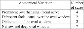
|
Facial nerve problems
Facial nerve was so prominent that stapedotomy could not be applied in one case and the operation was discontinued. The retraction of the nerve by using the suction probe as suggested by some authors is not practical and difficult to perform. In my opinion stopping the operation and suggesting a hearing aid, will be the most reliable decision in this case. Laser stapedotomy has been possible in the remaining 3 cases and 0.45 mm opening could be hardly performed to be able to insert a 0.4 mm piston prostheses. It will be difficult to try the stapedotomy by using needles or microdrill in these cases. The shaft of the microdrill may apply mechanical pressure and also give heat to the nerve causing postoperative facial nerve paralysis. Additionally in case of any complication regarding the footplate, it will be very difficult to manage the problem, as the vision will be very limited. With lasers we may need to reduce the spot size in these cases. For this purpose it will be necessary to get closer to the footplate. The close distance between the laser probe and the footplate may increase the heat effect in the vestibule. To reduce this, we suggest to keep intervals of 10 seconds between each laser shot.
Facial canal was dehiscent in 2 cases. Attention was required to preserve the nerve from the heat effect of laser in these cases. Insertion of a wet sponge over the nerve serves for keeping the nerve safe from the thermal effect of the laser.
Anatomical variations concerning
the oval window and footplate
Deeply situated narrow oval window is not uncommon. Generally it does not create any difficulty during the surgery. If the axis of the footplate does not permit direct access some difficulties may raise. Narrow and deep oval window was found in 7.7 % of primary stapedectomies. Thick footplate and obliterative otosclerosis were rare conditions and it was difficult to perform stapedotomy by means of conventional methods in these cases.
Perilymph gusher
Perilymph leakage has occurred after the first laser shot on the footplate. The flow rate was so high that the perilymph was filling the middle ear in seconds. The first measure was head elevation. Lumbar puncture and catheterization of the cerebrospinal fluid (CSF) was performed immediately. Following the decrease of CSF pressure, the leakage has stopped. A perichondrial graft was replaced over the footplate and teflon piston prosthesis has been inserted. In this case we observed that the footplate was quite thin and the crura were translucent as compared with the other cases. Following the surgery thiazide group diuretics are prescribed. The leakage continued for two days and decreased afterward. The bone conduction hearing levels were decreased step by step during the early postoperative period and total sensorineural deafness was developed in a week.
Fracture of the footplate
The footplate may be mobilized and/or fractured during the removal of the crura and opening of the vestibule. In case of rupture of the annular ligament floating footplate may occur. These complications likely occur while using mechanical techniques and its incidence is very low when using lasers. If the footplate is fractured and splitted in two pieces, the remaining part of the footplate is secured and the piston inserted into the vestibule by laying a fascia or perichondrium between the footplate and the piston to prevent leakage postoperatively, otherwise total stapedectomy may be the choice.
Findings in revision operations
Reparative granuloma
One case experienced neurosensorial hearing loss and vertigo a week following the primary operation. The tympanic membrane had a red color and bulged. The granuloma tissue was found to be filled the posterior mesotympanum and epitympanum. It surrounded the piston prosthesis as well as the ossicular chain. It was removed totally and the prosthesis has been reinserted, the middle ear has been packed with corticosteroid soaked gelfoam.
Incus problems
Erosion of the long process of the incus was the most frequent finding as a cause of late conductive hearing loss, in revision cases. It has been documented in 5 of 16 revision operations. Luxation of the ossicle is less frequent. Scar tissue and otosclerotic bone formation are other factors related with incus as being the cause of hearing loss.
Again malleus attachment piston prostheses are inserted in these cases to by-pass the eroded incus. The prosthesis is attached to the malleus as close to the processus brevis and the piston is inserted through the stapedotomy opening into the vestibule. It is difficult to see the malleus and the footplate with same angle while looking through the microscope. We advise to secure the wire to the manibrium of the malleus first and afterwards insert the piston into the vestibule.
Adhesive otitis media
Grade III retraction pocket formation was found in one case one year following the primary operation. The cholesteatoma was limited in the middle ear and the incus was eroded. The piston prosthesis has been extruded through the retracted membrane. Granulation tissue was developed beneath the retraction pocket. The retraction pocket was completely excised and the squamose epithelium was removed. The tympanic membrane was repaired by using composite perichondrium-cartilage graft and malleus attachment prosthesis was inserted.
Prosthesis dislocation
Dislocation of the prosthesis was observed in 6 cases. In all these cases the piston was not long enough to enter the vestibule for 1-1.5 mm. The length of the piston prostheses has to be prepared as it enters the vestibule for 1 to 1.5 mm. This is very important for a successful outcome. Shorter pistons usually migrate and the sound conduction mechanism fails. In these cases the prosthesis was found to be out of the vestibule. New piston prostheses were inserted to bridge the incus and the vestibule.
The remaining three cases had their revision surgery early in the postoperative period to explore the middle ear because of long prosthesis that caused vertigo in one case and in 2 cases, to evaluate the possibility of perilymph fistula, as one case experiencing early sensorineural hearing loss.
Discussion
With its high morbidity, the stapes surgery maintains being one of the most challenging operations of the ear. There is always a risk of creating floating footplate, dropping the bony fragments of the footplate into the vestibule while attempting to make the opening either by needles or microdrills. There is no doubt that the lasers are extremely effective and advantageous on creating the opening on the footplate for insertion of the prosthesis and it considerably decreased the incidence of complication rates. We are now aware that with laser stapedotomies, the incidence of sensorineural hearing loss, perilymph fistula occurrence has been reduced (1).
The major complications of stapedectomies rise during crurectomy and while creating the stapedotomy opening. Excision of the M. stapedius tendon by using micro scissors may also create mechanical trauma to the stapedial suprastructure. This may cause undesired mobilization of the stapes.
The visible lights of Argon and KTP lasers offer a great advantage on securing the structures such as facial nerve and inner ear structures. The wave length of Argon laser is 514 nanometers and KTP laser is 532 nanometers. Both lasers have similar properties. The only disadvantage that they have is the depth of penetration. By doing a rosette formation and keeping an interval of 10 seconds between each shoot secures the inner ear. The points on preferring the argon laser are: conduction of the beams by fiberoptic systems; control of capillary bleeding by means of photocoagulation; easier handling of the probes as they are similar to other microsurgical devices and control of the power by changing the working distance. The beam angle on tip of the probe is about 14 degrees and this decreases the power of the laser beams which enables a secure situation on protection of inner ear structures (2).
The anatomical variations that can not be documented preoperatively, should not disappoint the surgeon during the operation. Deeply situated narrow oval window is a common finding however it does not cause any problem in general. Additionally if the axis of the stapes is angulated, then working with the footplate becomes difficult especially when using conventional instruments.
Thick footplate and obliterative oval window are undesired conditions (3). The use of needles, perforators or the microdrills may increase the incidence of sensorineural hearing loss.
The position of the facial canal is very important during stapedotomy. If it is prominent and if it obscures the oval window, creating the opening on the footplate may be very difficult and risky. If the facial nerve overhangs prominently, then it may not be possible to perform the stapedotomy. In this condition some surgeons suggest retracting the nerve by using the suction tip or a needle. Even if the oval window is partially obstructed by the nerve, it may be difficult to create a safe stapedotomy by conventional techniques. The morbidity in this condition is two fold. In case of experiencing a slight problem on the foot plate, it may be impossible to manipulate safely. Serious complications such as floating footplate are more likely to occur in these conditions. Additionally, the efforts of creating a window on the foot plate can injure the facial nerve. When using lasers, the nerve has to be secured from the excessive heat created by laser. If the nerve is dehiscent, a wet sponge can successfully protect the nerve from the heat.
Tange and Bruijn mentioned that dehiscences were observed in 14 of the 427 patients (3.27%) who had a stapedotomy (4). This incidence is very low compared to the anatomical studies of the facial nerve in human cadaver temporal bones. Dehiscences of the facial canal are a variation of the normal anatomy of the facial nerve and these dehiscences occur sporadically in otosclerosis.
Ballester et al. observed in 6.7% of 595 stapedotomies the facial nerve had an abnormal course. And in one percent of all cases, there was a total prolapsus of the nerve over the oval window, with 2 special cases: facial nerve having an inferior course over the oval window and the promontory; facial nerve being widely spread over the oval window and the promontory. In patients with total facial nerve prolapsus, the prosthesis was either placed directly in a burr hole into the promontory just below the oval window (6 cases), or when the nerve ran over the promontory and over the oval window, the prosthesis was placed above the oval window at the site where the facial nerve is usually located (1 case) (5).
Gusher is a very rare phenomenon and it is generally associated with congenital stapes fixation or otosclerosis in adult age that may present during stapedectomy. A sudden perilymph flow occurs following stapedectomy, due to congenital malformation (abnormally wide cochlear aqueduct or internal auditory canal fistula), that causes an abnormal connection between subarachnoid and perilymphatic spaces (6). However this is not documented in every case. Our case with perilymph gusher exhibited pure conductive hearing loss and there we have not noted any sign of congenital malformation before the surgery. The only finding was the footplate was quite thin centrally and the crura were translucent. In spite of taking all the measures, sensorineural hearing loss was developed with in a week following the surgery.
Revision surgery should be regarded as a different concept than primary operations. The technique requires much more attention and the functional results are not as good as the primary operations. The decision for early revision operations generally are based on perilymphatic fistula, prosthesis dislocation and reparative granuloma formation.
One of the four cases that we performed early revision surgery were realized because of reparative granuloma. The granulomatous tissue was removed and the prosthesis was renewed in this case. Tange et al. indicated that reparative granuloma formation has been documented in 1.5% of cases which the gold prosthesis were used (7). This was figured as 329 reparative granuloma out of 319410 cases (incidence = 0.1%) in a poll study performed among the members of the American Otological Society and the American Neurotology Society (8). In this study 77 surgeons reported having encountered at least one reparative granuloma; and 50 surgeons related the occurrence of a reparative granuloma to a specific graft material. Gelfoam was the most common graft material reported when a reparative granuloma was encountered, followed by fat, and vertigo was the most frequently reported presenting symptom.
One of the most frequent findings in late revision cases is the erosion of long process of the incus. In stapes surgery, the attachment of the prosthesis to the long process of the incus plays an important role concerning the gain in hearing and the development of late complications such as incus erosion and necrosis. Band-shaped and spiral loops have been developed to achieve a broad, firm attachment to the long process of the incus. During stapes surgery, the view at the prosthesis is restricted, making it impossible to evaluate the effects of the differently shaped loops.
In a prospective study made by Lesinski 279 patients who referred for revision stapedectomy were analyzed (9). Prosthesis migration and subsequent fixation caused the majority (81%) of stapedectomy failures. Thirty-one percent had complete incus erosion, and additional 60% demonstrated partial incus erosion, usually on the undersurface of the incus. Residual fixed stapes footplate was found in 14%, and malleus fixation in 4%. Incus dislocation was found in 4%, and incus fixation in 2%.
The retraction pocket formation following stapedectomy is quite uncommon. Presumably it is associated with the negative pressure in the middle ear and decreased stiffness of the posterior-superior part of the tympanic membrane which could not resist to the negative pressure. The retraction pocket in this particular case was Grade III, with a squamose epithelium inside. The prosthesis was extruded and surrounded by the squamose epithelium. The long process of the incus was eroded. The retraction pocket and the squamose epithelium were completely removed, malleus attachment piston prosthesis was inserted and the tympanic membrane was repaired with cartilage graft. A similar case had been reported as developing bilateral cholesteatoma following successful stapedectomies by Ferguson et al. in 1986 (10).
The changes over the oval window that require revision surgery generally affects the hearing inversely, in contrast, revision surgery that is subjected to the ossicular chain problems have better postoperative hearing levels.
In conclusion, better surgical techniques are not always sufficient to receive good functional outcome from stapes surgeries, and the surgeon should also be aware of possible variations that affect the result of the surgery.
REFERENCES
Nissen RL. Argon laser in difficult stapedotomy cases. Laryngoscope 1998(Nov);108(11Pt1):1669-73.
Lundy LB. Otosclerosis update. Otolaryngologic clinics of North America 1996:29(2):257-63
Ayache D, Sleiman J, Tchuente AN, Elbaz P. Variations and incidents encountered during stapes surgery for otosclerosis. Ann Otolaryngol Chir Cervicofac 1999(Apr);116(1):8-14
Tange RA, de Bruijn AJ. Dehiscences of the horizontal segment of the facial canal in otosclerosis. ORL J Otorhinolaryngol Relat Spec 1997(Sep-Oct);59(5):277-9
Ballester M, Blaser B, Hausler R. Stapedotomy and anatomical variations of the facial nerve. Rev Laryngol Otol Rhinol (Bord) 2000;121(3):181-6
Cassano P, Decandia N, Cassano M, et al. Perilymphatic gusher in stapedectomy: demonstration of a fistula of internal auditory canal. Acta Otorhinolaryngol Ital 2003(Apr);23(2):116-9.
Tange RA, Schimanski G, van Lange JW, et al. Reparative granuloma seen in cases of gold piston implantation after stapes surgery for otosclerosis. Auris Nasus Larynx 2002(Jan);29(1):7-10.
Seicshnaydre MA, Sismanis A, Hughes GB. Update of reparative granuloma: survey of the American Otological Society and the American Neurotology Society. Am J Otol 1994(Mar);15(2):155-60.
Lesinski SG. Causes of conductive hearing loss after stapedectomy or stapedotomy: a prospective study of 279 consecutive surgical revisions. Otol Neurotol 2002(May);23(3):281-8.
Ferguson BJ, Gillespie CA, Kenan PD, Farmer JC Jr. Mechanisms of cholesteatoma formation following stapedectomy. Am J Otol 1986(Nov);7(6):420-4
|
Pro Otology |
Journal Home Contents Preview Next |