

|
Journal Home Contents Preview Next |
Pro Otology
Balkan Journal of Otology & Neuro-Otology, Vol. 4, No 1:57—61 © 2004
All rights reserved. Published by Pro Otology Association
Current Concepts for the Treatment of Midfacial and Frontal Skull Base Fractures
E. Zenev, Th. Guenzel, K.L. Bauchhage
Clinic of Ear, Nose and Throat Diseases, Central Clinic, Augsburg, Germany
ABSTRACT
Objective: Cars accidents remain major cause of severe midfacial and frontal skull base fractures.
Study design: The study design is a retrospective case review.
Setting: Clinic of Ear, Nose and Throat Diseases, Central Clinic – Augsburg.
Patients: 154 patients suffering from midfacial and skull base fractures have been treated for the period 2000-2002 year.
Interventions:
Results: From 2000 untl 2002 revisions of the frontal skull base with or without osteosynthesis in the midfacial area were performed on 154 patients. Local infections and postoperative bleedings made secondary surgery necessary in 5%. An osteomyelitis or removal of the osteosynthetic material due to inflammation did not occur in any case. On 6 patients a decompression of the optical nerve had to be performed. Two of these patients who were all blind on the affected side preoperatively had a satisfying view postoperatively. Two patients had only light perception and other 2 patients remained blind on the decompressed side.
Conclusion: In spite and because of the complex anatomy of the midface and fronral skull base the handling of trauma in this area is a challenge for the ENT surgeon. This is specialy important for the medical education of skull base surgeons, which should not be available only in few centers further on.
Key words: Skull base fractures, Midfacial fractures, Optical nerve decompression, Trauma.
Pro Otology 1:57—61, 2004
INTRODUCTION
As in the past car accidents remain major cause of severe injuries nowadays. In spite that, improvement of car design and introduction of airbags tend to decrease the incidence of skull base fractures. According to National Statistical department of Germany 362 054 car accidents happened in 2002 with dead toll of 6 842 victims. That was a decrease compared to 2000 when in 382 949 crashes died 7503 persons. According to Federal Service of Workers Protection and Occupational Medicine in the same year 1 170 000 domestic accidents had happened. 24.8% of the injuries were associated with bone fractures and 11.9% of them involved the head and facial region. Statistical estimation of indirect losses had revealed that there were average 32 lost days, and 15% of these days were in hospital ward.
In 2003 Federal Service of Workers Protection and Occupational Medicine have raised the slogan – “More safety for the children!”. Each year more than 300 000 sport and leisure time traumas were registered among children over 15 years old. In area of extreme sports such as budgie jumps, free climbing, deltaplaning and base jumping, traumas are less in number but much more severer in outcome.
In 1980 Richter, Brunner and Georgi have funded the basics of microsurgery and osteoplastic alternative techniques in patients with midfacial traumas and frontobasal cranial fractures with excellent results (2,4,15). We have had to admit that final operative results in some cases (wound and bone fracture healing) depends from the primary soft tissue damage. Development of functional deficits due to complex trauma, as well as deformations and psychosocial problems are not rare (2).
Anatomical features and classification of fractures
Brunner and Philkert showed us the proper attitude of bone thickenings at the area of middle facial region and the possibilities of osteoplastic manipulations. (FIG. 1, FIG. 2)(1).
In accordance of severity, area and direction of acting forces fractures can be divided in solitary, combined and complex. Le Fort and Escher Classifications are still useful and adequate in modern clinical evaluation.
Vogl for the firs time proposed a different approach in classification of fractures – according their radiological appearance (20). He has discriminated fractures of medial and lateral, isolated and combined fracture of the medial part of the face. As Brunner was mentioned fractures of medial parts of the face can be grouped as follows:
- Medial fracture – class I (FIG. 3) – without occlusial damage and can be managed by rhinosurgeons solitary;- Medial fracture – class II (FIG. 4) – with occlusial damage – the treatment here is provided by dentofacial surgeon solitary or in team with rhinosurgeon (1,2).
Isolated fractures of rhinobasis – Escher II can also be treated by rhinosurgeon following by secondary or endoscopic repair of dural rupture. All other types of fractures require neurosurgery.
|
|
Diagnostic and operative indications
The complexity of anatomical structures of orbit and orbital content explains the wide variety of traumatic injuries and combinations that are possible there especially fracture types involving intraorbital spaces. Telechantus and flattened of middle portion of the face are features easily coming on eye. The saving and supporting of vital functions of patients with dislocations of intracerebral spaces, opened cranial wounds combined with haemorrhagia and liquorrhea or lesions of eye bulb and optic nerve as well as all combined in polytrauma are at vigorous importance. (8,19,23).
Indications for optic nerve decompression often are unclear and complex. Recommendations in literature include conservative treatment, surgical approach or both. Traditional neurosurgical approaches for optical nerve decompression are craniotomy, extranasal transethmoidal approach or transantral microscopic controlled technique.
A cohort of 133 patients with traumatic optical nerve injuries were showed by Levin (12). His survey suggests that each pattern of therapy for every patient is chosen individually. Neither operative, neither conservative treatment with corticosteroids had proved definitely its own efficiency. Levin couldn’t say at last which combination is most appropriate. Wang and all. survey reaches similar conclusions (21).
The development of oedema depends mainly on primary care which patient with complex face and skull base fractures had received. It is very important to move forward primary care in the very beginning of accident at its first 7-8 hours even before face oedema occurred (8). Once oedema is fully advanced the operative intervention should wait for fourteen days. Later intervention however meets difficulties by advanced processes of bone healing.
|
||||||||
Material and methods
There were 2253 patients with facial and skull base fractures passed over to clinic of Ausburg in the period between 1998 and 2002. Approximately 60% of these were with fractures of nasal bones, orbital floor or zygomatic bone. All the results were nominated according the ISO-9 and ISO-10 Code score in data bank of the hospital. That’s why all the data, particularly after 2000 were estimated and analyzed. Data before 2000 are more at tentative character.
|
|
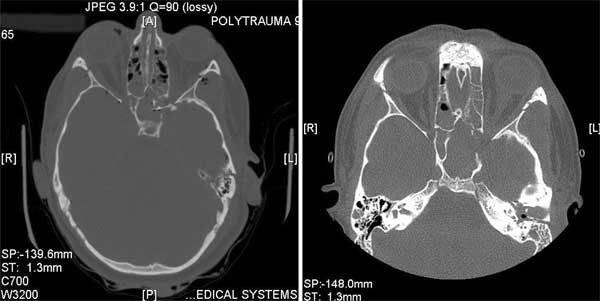
FIG. 5. Transseptal decompression of the optic nerve. |
Results
|
|
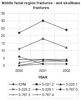
FIG. 6. ICPM–code classification alternative intervention in middle facial region with rhinobasis for 2000–2002. Legend: |
During these 3 years 154 patients were operated due to fractures of rhinobasis with or without osteosynthesis of the face middle portion. Partition with revisions of rhinobasis were estimated at 7,5%. Time distribution of these interventions are as follows: 2000 – 45 operations (2,2%); 2001 – 61 operations (3,0%); 2002 - 48 operations (2,3%). Microplates were used in 85% of patients. Miniplastic osteosynthetic materials were applied rarely (15%) and absorbable plates were applied even more rarely – in 5%. Among in one third of patients the interventions were followed by combined osteosynthesis with micro and miniplates.
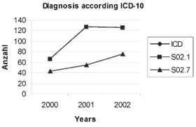
During 2000 twenty two patients (1,07%) went on frontobasal plastic of dura, 2001 – 30 patients (1,46%), 2002 – 24 patients (1,17%). Here we can detect a slight trend of increase. Conserved fascia lata and tissue glue were applied to close dura ruptures as a double patch among in 90% of patients. On figure 6 we can see ICPM – code classification alternative intervention in middle facial region with rhinobasis for 2000 – 2002. On FIG. 4 are shown patients with skull base fractures and those with complex fractures of medial part of the face for 2000 – 2002 as well. Here we can detect a slight trend of increase too.
For reconstruction of the frontal sinus anterior wall were used ear cartilage combined with titanium plate, bone dust and tissue glue. Laterally situated flaws were covered by neurosurgeons mainly with Palacos.
All surgical interventions have been performed under antibiotical coverage during the first week of intervention. Local infections and bleedings had led to revisions in less than 5% of cases. Such infections as osteomyelitis which could cause a removal of osteosynthesis had never been observed. In one patient we had observed a delayed type of contact allergic reaction of the skin laying immediately over the titanium osteosynthetic allograft. Skin lesion disappeared soon after removal of the titanium allograft which became possible after x-ray verification of good bone consolidation there.
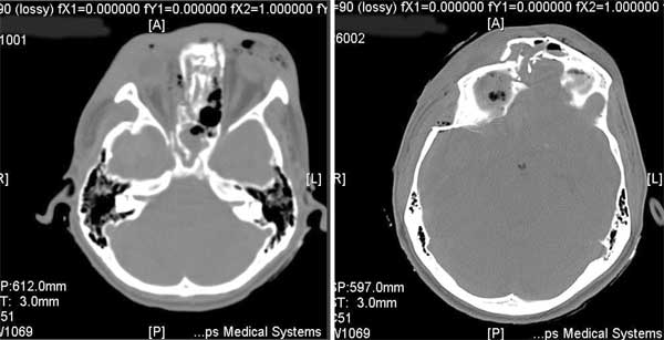
In 70% of patients natural formed traumatic lacerations were used as a surgical approach (Killian’s method, “glasses”) to deeper zones of skull damage. Figures 8 and 9 are examples of interdisciplinary cooperation.
A patient with complex fractures in the cerebral and facial parts of the skull and frontal skull base needs a primary neurosurgical craniotomy for alleviating of the intracranial pressure and for removal or reposition of bone fragments which compress optical nerve or internal carotid artery.
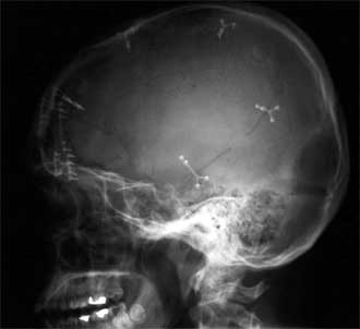
Two weeks after the first intervention another operation is have to be performed – second plastic of dura, cranialisation of frontal sinus cavity and osteosynthetic reconstruction of cranial and base parts of skull. Before the cranialisation to take place a scrutiny endoscopic removal of frontal sinus mucosa should be performed. Anterior wall of frontal sinus can be supported by autograft from epicranial aponeurosis, tissue glue and Tachocomb®. We realize osteosynthesis using microplates to restore lateroparietal bone defect with Palacos®. Antibiotical coverage is mandatory until 7th postoperative day. Rehabilitation of patient should start as soon as possible. If visual disturbance occurs carotid aneurism is suggested. We have had performed optic nerve decompression in 6 patient. In 5 patients we had decided to use transethmoidal – transsphenoidal approach through Killian’s section and in one patient (3 hours after the accident) a transseptal approach with removal of lateral walls of sphenoidal sinus was performed. In this case damaged eye had not light perception, but after optical nerve decompression it acquired visus of 0,5 ! (FIG. 5). Only in two patients blindness of the affected eyes remain unchanged after operations. Two other victims had reached light perception – from full blindness preoperatively.
Discussion
|
|
FIG. 7. Diagnosis according ICD 10. Legend: |
Primary care
When there aren’t proper soft tissue lacerations due to accident we use a corona like section of the skin. It ensures a good view and approach to fractures of whole upper and middle portions of facial skull (2,4,5,17,18). In case of already formed soft tissue lacerations in the middle portion of the face, we widen them most commonly in Killian’s pattern or as “glasses” type. We try to achieve such exposition of skull base that can allow us exact anatomical restoration and osteosynthesis of skull base (2-5). According Schick and all insufficient postrevisional cicatrisation of the dura without plastic closure means poor antibacterial defence (17). Dural lesions are life threatening thus their immediate closure is mandatory. Urgent operative intervention is obligatory in all cases of heavy rhinoliquorhhea with dramatic decrease of intracerebral pressure. The need of operation emerges also in already revealed symptoms of meningitis, worsening of the general condition and existing defect of dura. Postponing of operation within no more than 6 weeks is possible in asymptomatical liquorhhea or in meningitis that is susceptible to antibiotical therapy. According our consensus with Fulda Clinic every liquor fistula underpasses surgical closure (17,18). Small to middle defects of rhinobasis can be fixed with autogfafts from fascia lata or temporal muscle fascia and tissue glue with extradural transnasal approach with or without Tachocomb® plates (4,5,17). If there are broad destruction of posterior wall of the frontal sinus the most proper treatment represents cavity cranialisation with galleoperoistal flap. For frontal region reconstruction we use material from tabula externa (4,18). Adding of antibiotic in liquor fistula is not unusual. However there’s lack of statistical information under real efficiency of such prophylaxis. By the way evidences exists that there is no need of antibiotical usage among patients with small dural defects and lack of risk factors (diabetes mellitus, acute sinusitis, various immune suppressions) (22).
We have accepted the other authors principle of immediate optic nerve decompression with corticosteroid assistance (10,16,21).
Secondary care
For secondary reconstruction of traumatic defects, glasionomer cement from dentistry was used (4,5). Because of several lethal cases due to toxic reactions that had been reported in Belgium, France and Austria the usage of glasionomer cement was discontinued (9). The results with calcium phosphate cement in combination with titanium meshes are dubious (4,6,11,13).
Osteosynthesis
Since early 80’s many systems from different manufacturers were available. With the introduction of microplates and absorbable osteosynthetic materials one stage reconstruction became possible. Insufficient mechanical strength of absorbable osteosynthetic materials makes their application improper in zones with occlusial loadings (3,4,14,24).
|
|
FIG. 8. Complex skull fractures – cerebral, facial parts and skullbase. |
Conclusion
Advancement in passive and active defenses in car design decreased lethal outcomes but compensatory have had increased cases with severe and complex polytraumas. Certainly avoiding high speeds on road and extreme sports still remains the best prophylaxis to traumas.
Active treatment and maximum care from the very beginning of patient’s arrival combined with interdisciplinary approach are crucial factors to life support, successful treatment and good postoperative results.
|
|
FIG. 9. X-ray of skull after cranialisation and osteosynthesis. |
REFERENCES
Brunner FX, Kley W, Plinkert P. Anatomical studies and a correlative management of facial skeleton and skull base injuries with bone plate fixation Arch Otorhinolaryngol 1988;245:61-8.
Brunner FX. Surgical Management of Trauma Involving Skull Base and Paranasal Sinuses. In: Schmidek & Sweet. Operative Neurosurgical Techniques - Saunders, Philadelphia 2000:147-63.
Brunner FX. Osteosyntheseplatten im Gesichtsschadelbereich – ja oder nein? HNO 1995;43:205-8.
Brunner, FX. Klinik, Implantatmaterialien -was hat sich wo und wann bewahrt? European Archives of Oto-Rhino-Laryngology (Suppl) 1993;l.l:311-36.
Brunner FX, Geyer G, Hagen R. Primare und sekundare Rekonstruktionsmoglichkeiten van Gesichtsschadel- und Schadelbasisdefekten, Operationstechniken und Ergebnisse. ORL 1991;14:125-30.
Ducic Y. Midface reconstruction with titanium mesh and hydroxyapatite cement: a case report. Craniomaxillofac Trauma 1997;3(2):35-9.
Escher F. Das Schadelbasistrauma in oto-rhino-laryngologischer Sicht. HNO 1973;21:129-44.
Georgi W, Richter W, Brunner FX. Die Primarversorgung des zentralen, Mittelgesichtspfeilers mit stabiler Plattenosteosynthese. Laryng Rhinol Otol 1982;61:392–8.
Helms J, Geyer G. Alloplastic materials in skull base reconstruction. In Sekhar LN, Janecka IP (Eds). Surgery of cranial base tumors: A color atlas. New York: Raven Press, 1993:461-9.
Hong Y, Lin P, Liang Z. Clinical study of decompression of optic nerve through combined orbit, ethmoid and sphenoid approach. Lin Chuang Er Bi Yan Hou Ke Za Zhi 2001;15(12):546-9.
Kirschner RE, Karmachary J, Ong G, Gordon AD. Repair of the craniofacial skeleton with a calcium phosphate cement: quantitative assessment of craniofacial growth. Ann Plast Surg 2002;49(1):33-8.
Levin LA, Beck RW, Joseph MP. The treatment of traumatic optic neuropathy: the international optic nerve trauma study. Ophthalmology 2000;107(5):814.
Lucksanasombool P, Higgs RJED, Swain MV. Interfacial fracture toughness between bovine cortical bone. Biomaterials 2003;24(7):1159-66.
Richter W, Georgi W, Brunner FX. Die periorbitale, knöcherne Rekonstruktion (Miniplattenosteosynthese nach Champy). HNO 1982;30:186-8.
Samii M., Tatagiba M. Skull base trauma: diagnosis and management. Neurol Res 2002;24(2):147-56.
Shi J, Xu G, Xu J. Relationship between operative time and therapeutic effectiveness after the optic nerve injuries. Lin Chuang Er Bi Yan Hou Ke Za Zhi 2001;15(8):348-9.
Schick B, Weber R, Mosler P, et al. Langzeitergebnisse frontobasaler Duraplastiken. HNO 1997;45:117-22.
Schick B, Hendus J, Abd el Rahman el Tahan, Draf W. Rekonstruktion der Stirnregion mit Tabula extern a des Schadels. Laryngo-Rhino-Otol 1998;77:474-9.
Stoll W, Lübben B, Grenzebach U. Erweiterte Indikation zur Optikusdekompression: Eine differenzierte Analyse visueller Funktionseinschränkungen -auch bei bewusstlosen Patienten. Laryngo-Rhino-Otol 2001;80:78-84.
Vogi TJ, Balzer J, Mack M, Steger W. Radilogische Differentiladiagnostik in der Kopf-Hals-Region. Georg Thieme Verlag 1998:168-70.
Wang BH, Robertson BC, Girotto JA, et al. Traumatic optic neuropathy: a review of 61 patients. Plast Reconstr Surg 2001;107(7):1655-64.
Weber RK, Kaftan H, Draf W, Keerl R. Erste Erfahrungen mit der endonasalen Duraplastik ohne routine Gabe van Antibiotika. Laryngo-Rhino-Otol 2003;82:114-17.
Wohlrab TM, Maas S, de Carpentier JP. Surgical decompression in traumatic optic neuropathy. Acta Ophthalmol Scand. 2002;80(3):287-93.
Wiltfang J. Osteosynthesesysteme in der Mund-, Kiefer-, Gesichtschirurgie. HNO 2002;50:800-11.
|
Pro Otology |
Journal Home Contents Preview Next |