|
|
Journal Home Contents Preview Next |
Pro Otology
Balkan Journal of Otology & Neuro-Otology, Vol. 2, No 1:5-7 © 2002
All rights reserved. Published by Pro Otology Association
This new section for reporting recent articles is created
after the idea of Prof. Ugo Fisch, President of EAONO and
with the kindly permission of Prof. Robert Jackler, Editor of Otology & Neurotology.
The full text of the following article is published in
Otology & Neurotology, Vol. 22, No. 6, 2001.
Malleostapedotomy in Revision Surgery for Otosclerosis
*U. Fisch, †G. Acar, * †Al. Huber
*Ear, Nose and Throat Center, Hirslanden Clinic, Zurich, Switzerland
†Ear, Nose and Throat Department, Posta Telegraf Teskilati Education Hospital, Istanbul, Turkey
* †ENT Department, University Hospital Zurich, Switzerland
ABSTRACT
Purpose: The purpose of this study was to analyze the results of malleostapedotomy and to compare them with those of a conventional incus stapedotomy in a series of 82 consecutive surgical revisions in otosclerotic patients.
Materials and Methods: 82 consecutive revision stapes surgery cases over 5 years were evaluated. The preoperative and postoperative audiometric data of 80 (97.5%) of the patients were obtained.
Results: 71 of the patients underwent a functional revision procedure as malleostapedotomy (56.79%) or as incus stapedotomy (15.21%). The most common cause of failure of primary surgery was a displaced or malfunctioning prosthesis (86.2%). Pathologic changes of the oval window were found in 80% of the cases. Problems of the incus were identified in 80%and abnormality of the malleus in48.6% of the cases. The functional success rate of malleostapedotomy (closure within 10 dB) was found to be higher than that of traditional incus stapedotomy (p<0.05). Overclosure was seen in 12 patients (17%) and a significant sensorineural hearing loss in 2 patients (3%). There were no dead ears in this series. The postoperative hearing results after first revision surgery were better than those after multiple surgical procedures (p<0.005).
Conclusions: Malleostapedotomy yields better functional hearing results than incus stapedotomy in revision surgery for otosclerosis. The detection of many malleus fixations was the result of the systematic exposure of the anterior malleal process and ligament through an endaural approach with superior canaloplasty.
Key Words: Malleostapedotomy - Otosclerosis - Incus stapedotomy - Incus erosion - Otosclerosis.
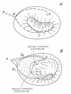
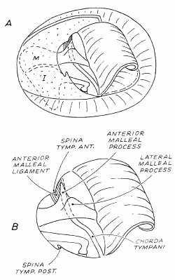
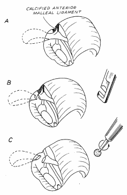
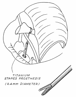
Malleostapedotomy (MS) is infrequently used in revision surgery for otosclerosis. Its main indications are severe incus erosion and malleus or incus fixation. The systematic use of the modified surgical technique used for revision procedures (an endaural approach combined with a superior canaloplasty) has permitted the discovery of numerous malleus and incus fixations that remained undetected at primary surgery. Hence, the number of malleostapedotomies performed in revision surgery for otosclerosis that increased in recent years. The aims of this article are to describe the surgical exposure necessary for adequate assessment of the mobility of the malleus and incus in revision surgery for otosclerosis, and to analyze the results of MS in relation to those of conventional incus stapedotomy (IS) in a series of 82 consecutive surgical revisions in otosclerotic patients (the performance of the operation follows the steps presented on figures 1, 2, 3, 4 and 5).
The described on Fig. 1, 2, 3 endaural approach with superior canaloplasty offers the advantage of direct visual control of the mobility of the anterior malleal process, the anterior malleal ligament, and the incudomalleal joint -essential for determination of partial fixation of the incus and the malleus.
The comparison of the mean preoperative and postoperative bone conduction thresholds, the air-bone gap (preoperative air minus postoperative bone conduction thresholds), postoperative air-bone gap for revision malleostapedotomy and incus stapedotomy (Table. 1), demonstrated a significant advantage of malleostapedotomy over incus stapedotomy.
The results of the study indicate that more attention should be paid to partial or total fixation of the malleus caused by calcification of the anterior malleal ligament particularly when conductive hearing loss persists after primary surgery.
REFERENCES
Han WW, Incesulu A, McKenna MJ, et al. Revision stapedotomy: intraoperative findings, results, and review of the literature. Laryngoscope 1997;107:1185-92.
Pederson CB. Revision surgery in otosclerosis: an investigation of the factors which influence the hearing result. Clin Otolaryngol 1996;21:385-8.
Fish U. Tympanoplasty, mastoidectomy and stapes surgery. Stuttgart, Germany: Georg Thieme Verlag, 1994:212-71.
Cokkeser Y, Naguib M, Aristegui M, et al. Revision stapes surgery: a critical evalution. Otolaryngol Head Neck Surg 1994;111:473-7.
Derlacki EL. Revision stapes surgery: problems with some solutions. Laryngoscope 1985;95:1047-53.
Glasscock ME, McKennan KX, Levine SC. Revision stapedectomy surgey. Otolaryngol Head Neck Surg 1987;96:141-8.
Hammerschlag PE, Fishman A, Scheer AA. A review of 308 cases of revision stapedotomy. Laryngoscope 1998;108:1794-800.
Langman AW, Lindeman RC. Revision stapedectomy. Laryngoscope 1993;103:954-8.
Deleted in proof.
Asai M, Roberson JB, Goode RL. Acoustic effect of malleus head removal and tensor tympani muscle section on middle ear reconstruction. Laryngoscope 1997;107:1217-22.
|
Pro Otology |
Journal Home Contents Preview Next |