
|
Journal Home Contents Preview Next |
Pro Otology
Balkan Journal of Otology & Neuro-Otology, Vol. 2, No 2:79-81 © 2002
All rights reserved. Published by Pro Otology Association
Malignant Skull Base Tumors in Childhood.
Case Report
*D. Marev, *R. Mutafova, *B. Banova, †V. Kaleva
*Department of Ear, Nose, Throat Diseases, †Department of Paediatrics Varna University of Medicine, Varna, Bulgaria
ABSTRACT
Objective: Skull base tumors in childhood present a complicated diagnostic and therapeutical problem because of its late clinical manifestations and poor prognosis.
Study design: In the period between 1997 and 2001 seven children 6 - 15 years of age were treated for malignant epipharyngeal neoplams at the Clinic of Ear, Nose, Throat Diseases and the IVth Paediatric Clinic.
Setting: A rare case is presented of osteoblastoclastoma of the sphenoidal bone body which is manifested in a way similar to that of malignant epipharengeal tumor.
Patient: The patient I.T.I. aged 15 felt sick at the end of December 1998 with sudden diplopia.
Intervention: Diagnostic, therapeutic. Main outcome measures: histological investigations of the tumor revealed malignant megaloblastic cellular tumor (osteoblastocalstoma).
Results: The diagnosis was confirmed by CAT - at the level of sella turcica a tumor formation of c. 6 cm in size was observed with a markedly expansive growth.
Conclusion: unilaterally impaired hearing of a conductive type with a headache of unknown aetiology, difficult nasal breathing and early cervical metastasis.
Key words: Skull base, Epipharynx, Tumor, Childhood.
Pro Otology 2: 79-81, 2002
INTRODUCTION
The tumor invasion through the skull base is a significant disease because it leads to serious complications. According to data from the National oncological centre the incidence of the malignant scull base tumors range around 0.56% of the overall population and 0.30% of all tumor localisations. In relation to the malignant neoplasms of the upper respiratory tract they occupy the second place among the carcinomas of the larynx. Regarding the histological forms, the first place in incidence is occupied by carcinoma - 50%, 15-16% of the cases are accounted by lymphoepithelioma, and mesenchymal tumors range between 30 - 40% of the cases. In childhood the undifferentiated spinocellular carcinoma prevails with peculiarities of lymphoepithelioma (1,4).
The nasopharyngeal carcinomas most commonly present with unilateral cervical adenopathy. The enlarged deep lymphatic node is typical for the upper posterior edge of the sternocleidomastoideus muscle. The lymphatic muscles become quickly enlarged and pronounced in the spinal accessoring chain and the posterior cervical triangle later involving the middle and lower jugular and supraclavicular nodes. The obstruction of the pharyngeal orificium of the Eustachian tube rather often results in serous otitis, e.g. the feeling of deafening of the ear, commonly accompanied by otalgia (2,3,7).
The tumor invasion through the skull base leads to the involvement of the skull and brain nerves. The lateral paralysis is also common. Blindness, entire ophthalmoplegia and acute pain in the inervated n. trigeminus regions are observed in the cases when the tumor invades the middle skulls pit and sinus cavernosus (6).
When the process spreads to the retroparotid region the cervical sympaticus becomes also invloved together with the IX to XII brain and skull nerves. They result in Horner's syndrome, difficulties swallowing, partial loss of taste, hemoanaesthesia of the soft palate, the pharynx and the larynx, dysphonia, unilateral paralysis of m. trapezius, m. sternocleidomastoideus or the tongue.
The posterior rhinoscopy revealed wide lesions in the region of the nasopharynx most commonly on its lateral side. It is often the case that the primary tumor is found by accident after the establishment of its haematogenic metastases in the lungs, the liver or the bones.
MATERIAL AND METHODS
In the period between 1997 and 2001 seven children aged 6 - 15 years were diagnosed and treated for malignant epipharyngeal formations at the Clinic of Ear, Nose, Throat Diseases and the Clinic of Paediatrcis at Varna University of Medicine. The clinical characteristics were determined by means of the subjective symptomatics, the tumor process localisation, stage specification according to TNM (the level of diagnostics), histomorphology and metastases.

|

|
|

|

|
|
|
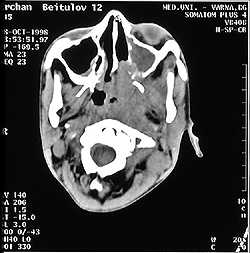
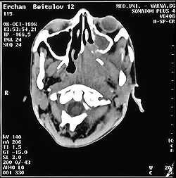
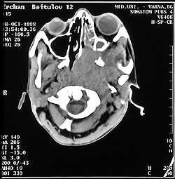
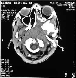
FIG. 1. CT scan - Tu formation with expansive growth. |
||
In 4 of the children the first manifested symptom was cervical adenopathy followed by unilateral nasal obstruction. Two children demonstrated a unilateral conductive impairment of hearing resulting from serous otitis in one of the patients and supurative otitis in the other. Recidivating epistaxes were observed in one of the children. In one of the cases we observed vision disturbances, e.g. diplopia, followed by headache, vomiting and a unilateral nasal obstruction.
The histomorphological structure revealed a predomination of the cases of undifferentiated spinocellular cancer with limphoepithlioma peculiarities (39.22%), followed by neurospinocellular cancer (37.25%) and hardening spinocellular cancer (11.70%).
Until the establishment of the diagnosis metastases were established in the regions of the lymphatic nodes in 70% of the children and 10% of the patients revealed metastases in distant organs which worsened the prognoses drastically. Our data were similar to those quoted by other authors (4,5). The early regional metastases found in 67% of the patients approximated the percentage reported by other authors.
A rare case of osteoblastoclastoma of the sphenoid bone body is presented and is manifested in a similar way as in a malignant tumor of the epipharynx.
The 15-year-old patient I.T.I. fell ill at the end of December 1998 with a sudden fit of diplopia. For about 2 months she was treated for Dg. Paresis nervia abducentis dex. and showed temporary improvement. In March 1999 she complained of severe headache in the occipital and the right frontal region followed by a "syphone" vomiting and almost entire right nasal obstruction. The posterior rhinoscopy revealed that the epipharynx was full of a whitish granulated Tu mass, easily bleeding at touch. The tumor was removed and investigated histologically (No. 29/05/05/99). The investigations revealed a malignant megaloblastic tumor (osteoblastoclastoma) most likely originating from the sphenoidal bone. In June the child lost her vision of the right eye completely and suffered discrete restrictions in the right eye bulbus movements in extreme abduction, anaesocoria with lack of direct pupil response to light. The eye fundi revealed a discrete retina oedema with slightly swollen venous vessels. The mesopharyngoscopy showed an immoble soft palate pushed downwards.
As a result of the histological findings and the clinical picture we decided that it was a case of an unusual localisation of the osteoblastoclastoma with a severely aggressive evolution toward the frontal and middle skull pit and the orbital contents. The diagnosis was confirmed by CAT at the level of the sella turcica. We could observe a large Tu formation sized about 6 cm in diameter which was well absorbing the contrastive materials and exhibiting a markedly expansive growth leading to destruction of the sella turcica and planum sphenoidale, more prominent in the epipharynx and constricting the epipharengeal space on the right (Fig. 1)
The general status and the blood picture did not show any deviations from the norm.
The administered treatment which brought about improvement included two cytostatic courses following the scheme: Vincristin, Farmorubicin, Endoxan.
CONCLUSIONS
1. The leading symptoms in malignant epipharyngeal tumors in childhood include unilaterally reduced hearing of a conductive nature, headache of unknown aetiology, difficult nasal breathing and early cervical metastases.
2. The cases which have been detected at a late stage are predominating - IIIrd - IVth stage (c. 73%).
3. Recidivating epistaxes are more commonly observed in children than in adults.
4. The most common histological form is the undifferentiated spinocellular carcinoma showing peculiarities of lymphoepithelioma.
REFERENCES
Boikikev Sv, Raichev R. Tumors of the upper respiratory tracts. Sofia: Medicina and Phyisics, 1979.
Deliiski M, Madjova V et all. Proteinuria and Hematuria in patients with Lung Cancer. Scr Scient Med 1992;28(1):125-6.
3. Dimov P. Methods and Diagnosis of the Desease. In: Chronic Otitis Media with Effusion. Stara Zagora: Ariel, 1997:28-35.
4. Kozlova A, Kalina O, Gamburg U. Tumors ORL. Mouskwa: Medicine 1979:80-1.
Malamov M, Georgiev G. Study of the ear-nose-throat tumors. Sofia: Medicine and physics 1983.
Paches A. Malignant tumors of the ORL. Mouskwa: Medicine 1988.
Stevens MN, A. Rich - Harisberger et al. A 10- year longitudinal study of tubal function in patients with nasopharyngeal carcinoma after irradiation. Arch. Otorhynolaringology 1975;3:735-9.
|
Pro Otology |
Journal Home Contents Preview Next |