|
|
Journal Home Contents Preview Next |
Pro Otology
Balkan Journal of Otology & Neuro-Otology, Vol. 2, No 2:77-78 © 2002
All rights reserved. Published by Pro Otology Association
Aneurysma Arteria Carotis Interna as Rare Entity in Otology.
Case Report
*Ang. Tudzarova, †Fr. Jakimovska, †S. Stojkova, †E. Dimeska, *S. Cvetanovska
*Skopje-Health Center, †Clinic of Otorhinolaryngology, Medical Faculty, Skopje, Macedonia
ABSTRACT
Objective: To present a patient with aneurysm of internal carotid artery as a very rare entity in otology, which is characterized with prolonged course and scarce symptomatology.
Study design: A rare retrospective review of the case.
Setting: The patient was referred to the Clinic of ORL where the necessary examinations and consultations were done and surgical treatment was indicated.
Patient: In December 2001, a 75-year old female came to our center complaining on tinnitus, buzzing and pain in the right ear as well as peritonsilar edema that caused difficulties in speech and swallowing.
Intrevention: Diagnostic, therapeutic (ORL examination, laboratory investigation, CT, MR, IA DSA- intra-arteial digital subtraction angiography, aneurysmectomy).
Main outcome measures: Giant aneurysm of a. carotis int. l. dex. with dimensions of 4x6 cm inframandibularily. Highly significant stenosis of 95% a. carotis int. l. dex.
Results: The results obtained have indicated that each symptom, apparently insignificant such as buzzing in the ears, if persists for a longer period of time and has certain characteristics should be examined in details so as to detect on time the cause for its onset.
Conclusions: The utilization of the known diagnostic methods such as CT, MRA, IA DSA.
Key words: Tinnitus, Ear, Aneurysm.
Pro Otology 2: 77-78, 2002
INTRODUCTION
Aneurysms of the extracranial carotid artery (ECA) are rare. Large single-institution series are seldom reported and usually are not aneurysm type-specific (1). Thus, information about immediate and long-term results of surgical therapy is sparse. Due to the fact that aneurysms are a rare phenomenon, studies concerning their examinations comprise a longer period of time (20, 30 years and even longer). Surgical therapy is the method of choice in the treatment of aneurysms.
OBJECTIVE
To present a rare case of aneurysma a. carotis int. l. dex., which was diagnosed on time and a successful aneurysmectomy was performed.
Aneurysms of carotid artery are uncommon, usually being associated with aneurysms at other sites and resulting from large-artery medial degeneration. They can be found in both extra and intracranial segment of the carotid artery. In this case aneurysm was situated in ECA.
MATERIAL
A female patient, aged 75. Two years ago she started to complain on a loud noise and buzzing in the right ear, most commonly manifested as pulsatile tinnitus. Hearing was not affected. Over the past two months, she had a pain in the right ear accompanied with increased intensity of the buzzing. Some disorders in swallowing and speech difficulties as well as stiffness on the right side of the face, also appeared.
METHODS
The methods which were used are ORL examination, Laboratory investigations, CT, MR angiography, IA DSA (intra-arterial digital subtraction angiography).
ORL examination. Peritonsilar edema on the right side suspected for peritonsilar abscess, was observed. The other ORL findings were OK. The incision revealed bloody fluids and therefore, it was suggested that additional investigations are necessary - CT, MR angiography and DSA.
Laboratory analyses. Preoperatively they did not significantly differ from the normal values: Le 7.7, Er 4.9, Hb 11.6, Hct 0.43, Tr 296, urea 8.3, creatinine 76.3, glycemia 7.0
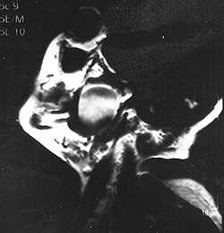
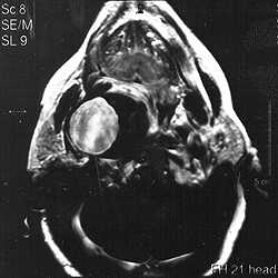
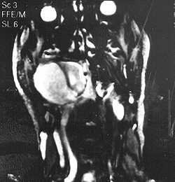
MR angiography. It has shown giant aneurysm of a. carotis int. l. dex. with dimensions 4x6 cm, situated inframandibularly and with high degree of occlusion (95%) that can be seen on the following figures: Fig. 1 - sagital plane, Fig. 2 - transverse plane, Fig. 3 - coronal plane.
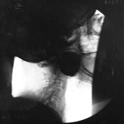
DSA. The results are presented in Fig. 4 and Fig. 5.
As soon as diagnosis was established, surgery was performed - aneurysmectomy of a. carotis int. l. dex and termino - terminal anastomosis with a. carotis int. There were no post-operative complications.
|
|
CONCLUSIONS
Cases like this are very rare in everyday ORl practice. Literature data report on cases with fatal outcome. In conclusion we could say that each symptom, although apparently insignificant, should carefully be examined since it could direct us to the right diagnosis. The utilization of the known diagnostic methods such as CT, MR angiography, IA DSA enable precise and rapid diagnosis.
REFERENCES
El Sabrout R, Cooley DA. Ekstracranial carotid artery aneurysms. J Vasc Surg 2000;31(4):702-12.
Zhang Q, Duan ZQ, Xin SJ et al. Management of extracranial carotid artery aneurysms. Eur J Vasc Endovasc Surg 1999;18(12):162-5.
Synowitz HJ, Leonhard T, Unger RR et al. Giant aneurysm - manifestations, diagnosis and therapy. Zentralbl Neurochir 1985;46(1):11-30.
Yasui T, Sakamoto H, Kishi H et al. Management of elderly patients with incidentally discovered unruptured aneurysms. No Shinkei Geka 1998;26(8):679-84.
|
Pro Otology |
Journal Home Contents Preview Next |