|
|
Journal Home Contents Preview Next |
Pro Otology
Balkan Journal of Otology & Neuro-Otology, Vol. 2, No 3:121-122 © 2002
All rights reserved. Published by Pro Otology Association
Cochlear Implantation in a Patient with Cogan’s Syndrome
and Ossified Cochlea. Case Report.
L. Migirov, T. Dagan, J. Kronenberg
Department of Otolaryngology Head and Neck Surgery, Sheba Medical Center, Tel
Hashomer and Tel Aviv University, Israel
ABSTRACT
Objective: Our goal was to review and discuss the implementation of cochlear implantation in Cogan’s Syndrome patients.
Study Design: Retrospective case review.
Setting: ENT department, Sheba Medical Center, Tel Hashomer, Israel.
Patients: Four patients with Cogan’s syndrome. One patient had an ossified cochlea.
Interventions: The patient with ossified cochlea underwent Suprameatal approach (SMA) cochlear implantation and the remaining three patients underwent posterior tympanotomy approach cochlear implantation.
Main Outcome Measures: The posterior tympanotomy and SMA were compared in order to assess the advantages and disadvantages of both in Cogan’s Syndrome patients and associated cochlear ossification.
Results: Four Cogan’s Syndrome patients were implanted; three had normal temporal bone anatomy and in the fourth patient ossification of the basal turn of the cochlea was demonstrated on high resolution CT scan. All patients were successfully implanted with no complications.
Conclusion: The SMA is a useful alternative to the conventional cochlear implantation technique in cases of ossified cochlea because of the wider exposure and safer entry into the middle ear it provides.
Key words: Cochlear implant, Cogan’s syndrome, Ossified cochlea.
Pro Otology 3: 121-122, 2002
INTRODUCTION
In 1945, Cogan described four patients with nonsyphilitic interstitial keratitis and audiovestibular dysfunction. This entity was later classified as Cogan’s syndrome (1). The pathophysiology of Cogan’s syndrome is now believed to be a generalized vasculitis with typical ophthalmological and histological findings. Audiological and vestibular manifestations of the disease include vertigo, tinnitus and rapidly progressive hearing loss (2).
The main observed histological change in the osseous and membranous labyrinth in Cogan’s syndrome patients is a process of new bone formation (2,3,4). Few cases of cochlear implantation in patients suffering from Cogan’s syndrome have been reported in the literature (5,6). Patients in these previous studies had normal temporal bone CT scans. Four patients with Cogan’s syndrome have been implanted in our department; three patients had normal temporal bone anatomy as demonstrated on high resolution CT scan and one patient had an ossified cochlea. In this study, we report our successful experience using the suprameatal approach (SMA) in cochlear implantation of the latter patient (7,8). Various techniques have been used for ossified cochlear implantation including facial recess approach with transcanal tympanotomy, radical mastoidectomy with total cochleostomy and middle cranial fossa approach (10,11,12,13). In comparison with other cochlear implantation techniques, the SMA proved to be a suitable alternative (7,8). It offered a wide exposure of the promontory and enables soft drill out of the cochlea in cases of cochlear ossification.
|
|
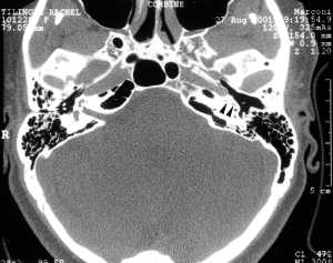
FIG 1. Ossification in basal turn of the cochlea (arrowheads). |
CASE REPORT
A 55-year-old female with a long history of progressive bilateral sensorineural hearing loss first presented with an acute onset of vertigo, left sided hearing loss and tinnitus at the age of 21. Over the next several years she had recurrent episodes of left sided hearing loss and vertigo. At age 25 she had an acute onset of bilateral hearing loss with ocular irritation. Her electronystagmography findings demonstrated bilateral areflexia. A serologic work-up for syphilis was negative and she was subsequently diagnosed with Cogan’s syndrome. Temporal bone CT scan revealed a partially ossified cochlear basal turn (figure). At age 53, a Nucleus 22 cochlear implant was inserted using the SMA technique (7,8). Ossification of the cochlea in this case required a drill out of the cochlear basal turn and the electrodes were inserted via the suprameatal tunnel into the cochleostomy. The postoperative course was uneventful. The patient had good hearing results as demonstrated in open set speech recognition scores one year following implantation.
DISCUSSION
Post mortem histological examination of temporal bones obtained from Cogan’s syndrome patients reveal ectopic bone formation in the perilymphatic spaces in the cochlear and vestibular end organs as well as ossification within the scala vestibuli of the basal turn. Other findings include cystic stria vascularis, edema of the spiral ligament, labyrinthine hydrops, degeneration of the organ of Corti, fibrosis within the perilymphatic cistern and macular destruction (2,3). Rarey et al demonstrated prominent ectopic osteogenesis in all the cochlear turns except the more basal scala tympani as well as in the semicircular canals (4). Casselman et al investigated two of six Cogan’s patients whose osteoneogenesis developed in juxtaposition to the round window and one case in which a bony narrowing appeared in the lateral semicircular canal (9).
Ossification of the cochlea in patients with Cogan’s syndrome may be detected on high resolution temporal bone CT. In the literature, patients suffering from Cogan’s syndrome have derived good to excellent results from the use of cochlear implants (5,6).
In our series, the initial three patients were operated before the development of the SMA technique. These patients had normal temporal bone CT scans and were operated using the posterior tympanotomy approach. However, in our experience with ossified cochlea prior to the fourth case, we found the keyhole view through the posterior tympanotomy to be less than satisfactory. We looked for a technique that would allow better visualization of the promontory and middle ear cavity in order to facilitate cochlear ‘drill out’.
CONCLUSION
The SMA provided a safe solution avoiding the risks of injury to the facial or chorda tympani nerves, middle cranial fossa dura and sigmoid sinus. Furthermore, it provided a route of entry similar to tympanoplasty in which the middle ear was widely exposed following elevation of a tympanomeatal flap and the cochleostomy site was thus best visualized.
Although further experience is required with the SMA technique in ossified cochlea, it seems to serve as a beneficial alternative for implanting ossified cochleas.
REFERENCES
Cogan DG. Syndrome of nonsyphilitic interstitial keratitis with vestibulocochlear symptoms. Arch Ophthalmol 1945;33:144-9.
Zechner GZ. Cogan syndrom. Acta Laryngol 1980;89:310-6.
Wolff D, Bernhard WG, Tsutsumi S, Ross IS, Nussbaum HE. The pathology of Cogan’s syndrome causing profound deafness. Ann Otol Rhinol Laryngol 1965;74:507-20.
Rarey KE, Bicknell JM, Davis LE. Intralabyrinthine osteogenesis in Cogan’s syndrome. Am J Otolaryngol 1986;4:387-90.
Cinamon U, Kronenberg J, Hildesheimer M, Taitelbaum R. Cochlear implantation in patients suffering from Cogan’s syndrome. J Laryngol Otol 1997;111:928-30.
Minet M, Deggouj N, Gersdorff M. Cochlear implantation in patients with Cogan’s syndrome: a review of four cases. Eur Arch Otorhinolaryngol 1997;234:259-62.
Kronenberg J, Migirov L, Dagan, T. Suprameatal approach: a new surgical approach for cochlear implantation. J Laryngol Otol 2001;115:283-5.
Kronenberg J, Migirov L, Dagan T. Suprameatal approach: a new surgical method for cochlear implantation. In: Jahnke K, Fischer M, et al. 4th European congress of Oto-Rhino-Laryngology Head and Neck Surgery, Germany. Monduzzi Editore, 2000:65-9.
Casselman JW, Majoor MHJM, Albers FW. MR of the inner ear in patients with Cogan’s syndrome. AJNR 1994;15:131-8.
Gantz BJ, McCabe BF, Tyler RS. Use of multichannel cochlear implants in obstructed and obliterated cochleas. Otolaryngol Head and Neck Surg 1988;98:72-81.
Montandon PB, Boex C, Pelizzone M. Ineraid cochlear implant in the ossified cochlea: surgical techniques and results. Am J Otolaryngol 1994;15:748-51.
Balkany T, Luntz M, Telischi FF, Hodges AV. Intact canal wall drill out procedure for implantation of totally ossified cochlea. Am J Otolaryngol 1997;18:58-9.
Colletti V, Fiorino FG. New window for cochlear implant insertion. Acta Otolaryngol 1999;119(2):214-8.
|
Pro Otology |
Journal Home Contents Preview Next |