
|
Journal Home Contents Preview Next |
Pro Otology
Balkan Journal of Otology & Neuro-Otology, Vol. 3, No 1:13-17 © 2003
All rights reserved. Published by Pro Otology Association
Tinnitus: Advances in Diagnosis and Management
Aristides Sismanis
Department of Otolaryngology - Head and Neck Surgery
Medical College of Virginia, Virginia Commonwealth University, Richmond, Virginia, USA
ABSTRACT
Objective: Tinnitus is a common otologic symptom secondary to numerous etiologies such as noise exposure, otitis, Meniere’s disease, otosclerosis, trauma, medications, and presbycusis. A thorough evaluation is necessary to rule out less common causes which may include acoustic neuromas, glomus tumors, atherosclerosis of the carotid arteries, arterioveous fistulae (AVFs) / malformations (AVMs) and intracranial hypertension.
Data sources: The literature used for the investigation is from the last 25 years.
Conclusion: Treating physicians need to have a very compassionate attitude towards these patients and statements such as “there is nothing that can be done” are very inappropriate and should be strongly condemned. Reassurance, hearing aids, masking devices, tinnitus retraining therapy, antidepressants, intratympanic medications and management of underlying pathologies such as carotid artery atherosclerosis, skull base tumors, intracranial hypertension and AVMs/AVFs provide relief for the majority of these patients.
Key words: Tinnitus, Diagnosis, Management.
Pro Otology 1: 13-17, 2003
INTRODUCTION
Tinnitus is a common otologic symptom secondary to numerous etiologies. Based upon sound characteristics, tinnitus can be classified as non-pulsatile (or continuous) which is the most common type and pulsatile. Since these two types of tinnitus have very diverse etiologies, pathophysiology as well as treatment, they will be described as separate entities.
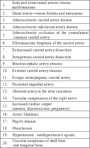
| Table 4. Arterial etiologies of pulsatile tinnitus. |

|
II. PULSATILE TINNITUS
Pathophysiology and Classification
Pulsatile tinnitus (PT) originates from sounds produced by vascular structures within the cranial cavity, head and neck region and thoracic cavity, which are transmitted to the cochlea by bony and vascular structures. Pulsatile tinnitus arises from either increased flow volume or stenosis of a vascular lumen. Vascular PT can be classified as arterial or venous according to the vessel of origin. Venous PT type can originate not only from primary venous pathologies, but also from conditions causing increased intracranial pressure by transmission of arterial pulsations to the dural venous sinuses (35). Pulsatile tinnitus originating form other non-arterial structures is classified as non-vascular. Pulsatile tinnitus can be called objective or subjective according to whether it is audible to both the patient and examiner or to the patient only.
ARTERIAL ETIOLOGIES
Atherosclerotic Carotid Artery Disease
Atherosclerotic carotid artery disease (ACAD) is a common cause of PT in older than 50 years of age patients, especially when associated risk factors for atherosclerosis such as hypertension, angina, hyperlipidemia, diabetes mellitus, and smoking are present. Objective PT can be the first manifestation of ACAD in some of these patients (36). Pulsatile tinnitus in ACAD is secondary to bruit(s) produced by turbulent blood flow at stenotic segment(s) of the carotid artery. Diagnosis can be confirmed by Duplex Ultrasound Studies (36). Table 4 summarizes the arterial etiologies. (37).
VENOUS ETIOLOGIES
Pseudotumor cerebri syndrome
Pseudotumor cerebri syndrome is a common cause of venous PT, especially in young and obese females. This syndrome is characterized by increased intracranial pressure without focal signs for neurological dysfunction except for occasional fifth, sixth, and seventh cranial nerve palsies. Diagnosis of this syndrome is made by exclusion of lesions producing intracranial hypertension. Although the classic presentation of pseudotumor cerebri syndrome consists of headaches and/or visual disturbances, PT alone or in association with hearing loss, dizziness and aural fullness has been reported as the main manifestation(s) of this syndrome (35). Many of these patients are morbidly obese and have associated papilledema. Absence of papilledema, however, does not exclude this entity (38). Head CT or MRI is normal in the majority of patients, although an empty sella and/or small ventricles may be present. Diagnosis should be established by lumbar puncture and confirmation cerebrospinal pressure (CSF) of more than 200 mm of water with normal CSF constituents.
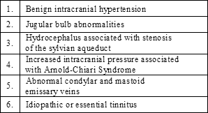
| Table 5. Venous etiology of pulsatile tinnitus. |

|
Pulsatile tinnitus in pseudotumor cerebri syndrome is believed to result from the systolic pulsations of the CSF originating mainly from the arteries of the Circle of Willis. These pulsations, which are increased in magnitude in the presence of intracranial hypertension, are transmitted to the exposed medial aspect of the dural venous sinuses (transverse and sigmoid) compressing their walls synchronously with the arterial pulsations. The periodic compressions of the dural sinuses convert the normal laminar blood flow to turbulent, thus producing a low frequency PT (35). The low frequency sensorineural hearing loss seen in many of these patients is believed to result from the masking effect of the PT. This is supported by the fact that in most of these patients light digital compression over the ipsilateral internal jugular vein (IJV) results in cessation of the tinnitus and immediate improvement or normalization of hearing (35). Stretching or compression of the cochlear nerve and brain stem and/or edema, can also play a role in the hearing loss and dizziness encountered in these patients. This is supported by the abnormal auditory evoked responses present in one third of these patients (39).
Idiopathic or Essential Pulsatile Tinnitus
Idiopathic PT, essential PT and venous hum are terms used interchangeably to describe patients with PT of unclear etiology. A possible cause of idiopathic PT is believed to be turbulent blood flow produced in the IJV as it curves around the lateral process of the atlas (40). Diagnosis of this condition should be made only after appropriate evaluation and elimination of other disorders, such as pseudotumor cerebri syndrome. Table 5 summarizes the venous etiologies of PT (37).
NON-VASCULAR ETIOLOGIES
Palatal, Stapedial, and Tensor Tympani Muscle Myoclonus
Myoclonic contractions of the tensor veli palatini, levator veli palatini, salpingopharyngeus and superior constrictor muscles can result in objective PT. These contractions can range between 10 and 240 per minute and may be confused with the arterial pulse. This disorder is often seen in younger patients, usually within the first 3 decades of life, although it may be seen in older individuals as well (41). Associated neurologic disorders such as brain stem infarctions, multiple sclerosis, trauma, and syphilis have also been reported. Involvement of the olivary tracts, posterior longitudinal bundle, dentate nucleus, and reticular formation has been identified in these patients (41).
Myoclonus of the stapedial muscle has also been reported as a cause of pulsatile tinnitus (42).
EVALUATION
History
Associated symptoms of hearing loss, aural fullness, dizziness, headaches and visual disturbances, such as visual loss, transient visual obscurations, retrobulbar pain and diplopia are suggestive of associated Pseudotumor cerebri syndrome (35,37).
Older patients with history of cerebrovascular accident, transient ischemic attacks, hyperlipidemia, hypertension, diabetes mellitus and smoking should be suspected of having ACAD (36). Females with associated headaches, dizzy spells, syncope, fatigue, and lateralizing neurological deficits should be evaluated for fibromuscular dysplasia (FMD) (43). Sudden onset of PT in association with cervical or facial pain, headache, and symptoms of cerebral ischemia is highly suggestive of extracranial or intrapetrous carotid artery dissection (44).
Examination
Otoscopy is essential for the detection of middle ear pathology such as a high or exposed jugular bulb, aberrant carotid artery, and glomus tumor. Rhythmic movements of the tympanic membrane can be present in patients with tensor tympani myoclonus.
Head and neck examination is very important. A palpable cervical thrill is suggestive of a cervical arterio-venous malformation. Myoclonic contractions of the soft palate can be present in patients with palatal myoclonus. Wide opening of the oral cavity during examination may result in elimination of the soft palate contractions.
Auscultation of the ear canal, peri-auricular region, orbits, cervical region and chest is essential for detection of objective PT, bruits and heart murmurs. This should be performed in a quiet room, preferably with a modified electronic stethoscope. Auscultation with an electronic stethoscope has been found more sensitive than traditional auscultation (45). When objective PT is detected, its rate should be compared to the patient’s arterial pulse. The effect of light digital pressure over the ipsilateral IJV should be determined. Pulsatile tinnitus of venous origin, as in pseudotumor cerebri syndrome, decreases or completely subsides with this maneuver (35). In patients with the arterial type of PT, however, this maneuver is ineffective in decreasing the intensity of tinnitus. A complete neurologic examination, including funduscopy, should be included in the examination. Papilledema is compatible with pseudotumor cerebri syndrome, its absence, however, does not exclude this entity (38).
Audiologic Testing
Pure tone (air and bone conduction) and speech audiometry should be performed in all patients.
Impedance audiometry can be helpful in patients suspected of tensor tympani myoclonus.
Metabolic Workup
Complete blood count and thyroid function tests should be obtained in patients with increased cardiac output syndrome to exclude anemia and hyperthyroidism. Serum lipid profile and fasting blood sugar should be requested in patients suspicious for ACAD.
Ultrasound Studies
Duplex carotid ultrasound (including the subclavian arteries) and echocardiogram should be obtained in patients suspected of ACAD and valvular disease respectively (35).
RADIOLOGIC EVALUATION
Radiologic evaluation needs to be individualized according to the clinical presentation, physical and audiometric findings. The following represent the author’s radiologic evaluation protocol:
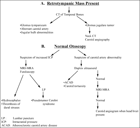
1. For patients with normal otoscopic findings, screening with high quality magnetic resonance angiography (MRA)/magnetic resonance venography (MRV) in conjunction with brain MRI, should be initially obtained. In patients with objective PT and/or a head bruit whose MRI/MRA/MRV studies are normal, carotid angiography should be strongly considered to exclude small dural AVMs/AVFs (46).
2. Patients with a retrotympanic mass should have a high-resolution temporal bone computed tomography (CT) at their initial evaluation (47). If a glomus tympanicum, aberrant internal carotid artery, or jugular bulb abnormality is diagnosed, no other imaging studies are needed. For patients with glomus jugulare tumors, CT examination of the neck also should be obtained to detect any possible synchronous chemodectomas along the carotid arteries. Carotid angiography is indicated only for prospective surgical cases to evaluate the collateral circulation of the brain (arterial and venous) in anticipation of possible vessel ligation and/or preoperative tumor embolization. FIG. 1 depicts an algorithm for evaluating PT patients.
MANAGEMENT
The following describe treatment modalities for the most common etiologies of PT.
Pseudotumor cerebri syndrome patients should be treated for any associated disorders such as morbid obesity. These patients responds well to weight reduction and medical management with Acetazolamide (Diamox). Weight reduction surgery was found effective in relieving PT in 13 out of 16 morbidly obese patients with associated pseudotumor cerebri syndrome who had failed conservative management (48).
Symptomatic high-dehisced jugular bulb can be repaired with pieces of cortical bone or cartilage (37). Pulsatile tinnitus secondary to otosclerosis may respond to stapedectomy (37). Botulinum toxin has been used successfully for treating palatal myoclonus (49). Tensor tympani and stapedial myoclonus may respond to section of the respective muscles via tympanotomy (42). Pulsatile tinnitus secondary to the antihypertensive medications enalapril maleate or verapanil hydrochloride subsides soon after discontinuation of these agents (37). Dural AMVs and AVFs are treated in the majority of patients with selective embolization. Vascular neoplasms can be treated surgically or with stereotactic radiotherapy.
|
FIG 1. Pulsatile tinnitus diagnostic algorithm. |

|
Ligation of the ipsilateral to the tinnitus IJV has been recommended in the literature for patients with idiopathic PT. The results of this procedure, however, have been very inconsistent and poor overall (40). It is strongly recommended that this procedure be considered only after careful elimination of any other causes of PT, especially pseudotumor cerebri syndrome. The author remains very skeptical regarding the value of this procedure for any patient.
CONCLUSION
Recent advances in tinnitus research have led to a better understanding and management of this common otologic symptom. A thorough evaluation should be performed in all patients in order to accomplish accurate diagnosis and effective management.
In the majority of patients explanation and reassurance are very effective. Hearing aids, masking devices, retraining methods, antidepressants, intratympanic medications and management of underlying pathologies such as carotid artery atherosclerosis, skull base tumors, intracranial hypertension and AVMs/AVFs are very helpful in the majority of these patients.
REFERENCES
Vernon J: Tinnitus causes, evaluation and treatment in otolaryngology (ed) G.M. English. Philadelphia: Lippincott Company, 1992.
Vernon J: Tinnitus 1989: current knowledge and treatment therapy. The Hearing Journal 1989;42(11):7-11.
National Center for Health Statistics: Basic data on hearing levels of adults 25-74 years, United States 1971-1975. Vital and Health Statistics Publication Series 11, No. 215. Hyattsville, Maryland, US Dept of Health Education and Welfare, 1980.
Schleuning A: Medical aspects of tinnitus. The Hearing Journal 1989;42(11):12-5.
Dobie RA: Tinnitus and depression. The International Tinnitus Journal 1997;3(1):33-4.
Moller AR. Similarities between chronic pain and tinnitus. American Journal of Otolaryngology 1997;18:577-85.
Jastreboff PJ, Gray WC, Gould SL. Neurophysiological approach to tinnitus patients. American Journal of Otolaryngology 1996;17:236-40.
Schleuning AJ, Johnson RM. Use of Masking for Tinnitus. International Tinnitus Journal 1997;3(1):25-9.
Jastreboff PJ, Jastreboff MM. Tinnitus Retraining Therapy (TRT) as a method for treatment of tinnitus and hyperacusis patients. Journal of the American Academy of Audiology 2000;11(3):162-77.
Steenerson RL, Cronin GW. Treatment of tinnitus with electrical stimulation. Otolaryngology Head and Neck Surgery 1999;121(5):511-3.
Aschendorff A, Pabst G, Klenzner T, et al. Tinnitus in Cochlear Implant Users: The Freiburg Experience. The International Tinnitus Journal 1998;4(2):162-4.
Sullivan M, Katon W, Russo J. A randomized trial of nortriptyline for severe chronic tinnitus. Archives of Internal Medicine 1993;153:2251-9.
Israel JM, Connelly JS, McTigue ST, et al. Lidocaine in the treatment of Tinnitus Aurium. Archives of Otolaryngology 1982;108:471-3.
Coles RR, Thompson AC, O’Donoghue GM. Intra-tympanic injections in the treatment of Tinnitus. Clinical Otolaryngology 1992;17(3):240-2.
Podoshin L, Fradis M, David YB. Treatment of tinnitus by intratympanic instillation of lignocaine (lidocaine) 2 per cent through ventilation tubes. Journal of Laryngology and Otology 1992;106(7):603-6.
Rudack C, Hillebrandt M, Wagenmann M, et al. Treatment of tinnitus with lidocaine? A report of clinical experiences. HNO 1997;45(2):69-73.
Sanchez TG, Balbani AP, Bittar RS, et al. Lidocaine test in patients with tinnitus: rationale of accomplishment and relation to the treatment with carbamazepine. Auris Nasus Larynx 1999;26(4):411-7.
Rosenberg SI, Silverstein H, Rowan PT, et al. The effect of melatonin tinnitus. Laryngoscope 1998;108(3):305-10.
Johnson RM, Brummett R, Schleuning A. Use of Alprazolam for relief of tinnitus: A double-blind study. Archives of Otolaryngology Head & Neck Surgery 1993;119:842-5.
Kaasinen S, et al. Effect of intratympanically administered gentamycin on hearing and tinnitus in Ménière’s disease. Acta Otolaryngol (Stockh) Suppl 1995;520:184-5.
Sakata E, Ito Y, Itoh A. Clinical Experiences of Steroid Targeting Therapy to Inner Ear for Control of Tinnitus. International Tinnitus Journal 1997;3(2):117-21.
Davies E, Knox E, Donaldson I. The usefulness of nimodipine, an L-calcium channel antagonist, in the treatment of tinnitus. Br J Audio 1994;28:125-9.
Moller AR. Tinnitus. In: Jackler RK, Brackmann DE, eds. Neurotology. St. Louis: Mosby-Year Book, 1994;153-65.
Pulec JL. Tinnitus: surgical therapy. American Journal Otology 1984;5:479-80.
Fisch J. Surgical treatment of vertigo. Journal of Laryngology and Otology 1976;90:75-86.
Silverstein H: Transmental Labyrinthectomy with and without cochleovestibular neurectomy. Laryngoscope 1976;86:1777-91.
House JW, Brackmann DE. Tinnitus, Surgical Management. Ciba Found Symp 1981;85:204-16.
Gardner G. Neurologic Surgery and tinnitus. Journal of Otolaryngology (Suppl) 1984;9:311-8.
Barrs DM, Brackmann DE. Translabyrinthine nerve section: Effect of tinnitus. Journal of Laryngology and Otology 1984;9:287-93.
Silverstein H, Haberkamp T, Smouha E. The state of tinnitus after inner ear surgery. Otolaryngology Head and Neck Surgery 1986;95:438-41.
Glasscock MC, Thedinger BA, Cueva RA. An analysis of the retrolabyrinthine vs the retrosigmoid vestibular nerve section. Otolaryngology Head and Neck Surgery 1991;104:88-95.
Wazen JJ, Caruso M. Tinnitus outcome in surgery for vertigo. International Tinnitus Journal 1997;3(1):51-3.
Okamura T, Kurokawa Y, Ikeda N, et al. Microvascular decompression for cochlear symptoms Journal of Neurosurgery 2000;93(3):421-6.
Gersdorff M, Nouwen J, Gilain C, et al. Tinnitus and otosclerosis. Eur Arch Otorhinolaryngol 2000;257(6):314-6.
Sismanis A. Otologic manifestations of benign intracranial hypertension syndrome: diagnosis and management. Laryngoscope 1987;42(97):1-17.
Sismanis A, Stamm MA, Sobel M. Objective tinnitus in patients with atherosclerotic carotid artery disease. American Journal of Otology 1994;15:404-7.
Sismanis A. Pulsatile tinnitus. A 15-year experience. American Journal of Otology 1998;19(4):472-7.
Spence JD, Amacher AL, Willis NR. Benign intracranial hypertension without papilledema: Role of 24-hour cerebrospinal fluid pressure. Neurosurgery 1980;7(4):326-36
Sismanis A, Callari RH, Slomka WS, Butts FM: Auditory evoked responses in benign intracranial hypertension syndrome. Laryngoscope 1990;100:1152-5.
Hentzer E. Objective tinnitus of vascular type. Acta Otolaryngol 1968;66:273-81.
Heller MF. Vibratory tinnitus and palatal myoclonus. Acta Otolaryngol Stockh) 1962;55:292-8.
Zipfel TE, Kaza SR, Greene JS. Middle-ear myoclonus. Journal of Laryngology and Otology 2000;114(3):207-9
Wells RP, Smith RR. Fibromuscular dysplasia of the internal carotid artery: a long follow-up. Neurosurgery 1982;10:39-43.
Saeed SR, Hinton AE, Ramsden RT, et al. Spontaneous dissection of the intrapetrous internal carotid artery. Journal of Laryngology and Otology 1990;104:491-3.
Sismanis A, Butts, FM. A practical device for detection and recording of objective tinnitus. Otolaryngology Head and Neck Surgery 1994;110:459-62.
Shin EJ, Lalwani AK, Dowd CF. Role of Angiography in the Evaluation of Patients with Pulsatile Tinnitus. Laryngoscope 2000;110:1916-20.
Sismanis A, Smoker WRK. Pulsatile Tinnitus: Recent Advances in Diagnosis. Laryngoscope 1994;104:681-8.
Michaelides EM, Sismanis A, Sugerman HJ, et al.: Pulsatile tinnitus in patients with morbid obesity: the effectiveness of weight reduction surgery. American Journal of Otology 2000;21(5):682.
Jero J, Salmi T. Palatal myoclonus and clicking tinnitus in a 12-year-old girl - case report. Acta Otolaryngol Suppl 2000;543:61-2.
Sismanis A, Butts FM, Hughes GB. Objective tinnitus in benign intracranial hypertension: an update. Laryngoscope 1990;100:33-6.
|
Pro Otology |
Journal Home Contents Preview Next |