
|
Journal Home Contents Preview Next |
Pro Otology
Balkan Journal of Otology & Neuro-Otology, Vol. 4, No 2-3:101103 © 2004
All rights reserved. Published by Pro Otology Association
Differential Diagnosis in Hearing Loss
D. Popova, J. Spiridonova, S. Varbanova
ENT Department, University Hospital, Sofia, Bulgaria
ABSTRACT
Objective: The aim of this study is to describe hearing disorder in witch sound enters the inner ear normally but the transmission of signal from inner ear to the brain is impaired. The patients display auditory characteristics consistent with normal outer hair cell function and abnormal neural function at the level of the VIII-th nerve or the damage of inner hair cells. The typical profile of Auditory neuropathy is a sensorineural hearing loss (often low frequency), poor speech perception, absent or severely abnormal ABR, CM present, OAE present, no acoustic reflex (ipsi at contra lateral).
Setting: ENT Department, University Hospital, Sofia, Bulgaria.
Conclusion: Problems in differentiation of neural vs. sensory deafness during screening; the role of OAE in the hearing impaired (lee 2001); the diagnosis of AN should not be a immediate referral for CI. Improved or fluctuating auditory responses with increasing ages are possible.
Key words: Auditory neuropathy, Hearing loss, ABR, CM, OAE.
Pro Otology 2-3:101103, 2004
Introduction
Hearing is the transduction of sound (mechanical energy) into neural impulses and the interpretation of those impulses by the central nervous system. Hearing loss can result from a defect at any level in this system. They must be:
conductive hearing loss:
external ear; middle ear;
sensorineural hearing loss.
Often there is poor discrimination out of proportion to degree of pure tone sensitivity, due to distortion of sound by cochlear to nerve.
The basic parameters of hearing loss are:
1. Anomalies of intensity coding loudness is a function of number of auditory nerve fibers firing and their discharge rate;
2. Anomalies of frequency coding:
place coding hair cells at maximal displacement of basilar membrane are maximally stimulated;
volley coding hair cells fire at same frequency as sound;
telephone place coding currently most popular theory, low frequency sounds are volley coded; high frequency place coding and at mid frequency, both mechanisms are operative.
3. Impulses transmitted to brain via acoustic nerve with projections to both sides;
4. Central perception and interpretation.
What is auditory neuropathy:
Auditory neuropathy is a hearing disorder in witch sound enters the inner ear normally but the transmission of signal from inner ear to the brain is impaired. It can affect people of all ages from infancy through adult hood. The number of people affected by auditory neuropathy is not known, but the condition affects a relatively small percentage of people who a deaf or hearing impaired.
People with AN have poor speech perception abilities, they have trouble understanding speech clearly, often speech perception is worse than would be predicted by the degree of hearing loss. They are able to hear sounds but have difficulty recognizing spoken words. Sounds may fade in and out for these individuals and seem out of sync.
The patients displays auditory characteristics consistent with normal outer hair cell function and abnormal neural function at the level of the VIII-th nerve or the damage of inner hair cells
Auditory neuropathy typical profile:
hearing loss- sensorineural often low frequency;
poor speech perception;
absent or severly abnormal ABR;
CM present;
OAE present;
no acoustic reflex (ipsi at contra lateral).
The AN is not a new hearing disorder, a new is our possibility to clinical identify the disorder and distinguish it from other problems. That has become possible primarily because of clinical use of otoaoustic emissions in recent years.
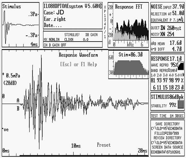
EOAEs are believed to be generated by electromotile activity of the outer hair cells of the cochlea and they represent a preneural phenomenom. The clinical implication is non invasive direct window into the cochlea.

|
|
FIG. 1. Graphics of EOAEs. |
EOAEs intended to discriminate between cochlear and neural functioning and assist in differential diagnosis of auditory disorders. The outer hair cells are implicated in the fine tuning of cochlear micromechanics and the underlying nonlinear processes. They are of interest of their putative contribution to the sensitivity and frequency selectivity of the hearing organ.
Many reports have suggested that EOAEs may be produced and measured independent of the neural integrate of the auditory system. Norton (1992) wrote It is important to remember when applying OAEs to clinical populations that their generation is preneural and independent of both afferent and efferent innervation. That is, if a lesion is central to outer hair cells, OAEs could be present and behavioral and neural responses depressed.
The pattern of normal outer hair cells function combined with abnormal neural responses shown by ABR places the site of AN to the area including the inner hair cells, the tectorial membrane, and the cochlear branch of the VIII-th cranial nerve, the VIII-th nerve itself and perhaps auditory pathways of the brainstem et etc. Neural problems may be axonal or demyelinating. Afferent as well. as efferent pathways may be involved.
The clinical auditory tests which must use can be sensitive to cochlear and auditory nerve function.
OHC function can be evaluated be measuring OAEs and cochlear microphonics. Clinical tests that are specifically sensitive to auditory nerve dysfunction are middle ear reflexes (ipsi and contra lateral), ABR and pure tone audiogramm and word recognitions tests in noise.
OAEs are related to the mechanical function of the outer hair cell system. This mechanism is important, but not insufficient. Inner hair cell function is also necessary to activate the sensory processus that transmit incoming information to the auditory nerve and central auditory sytem.
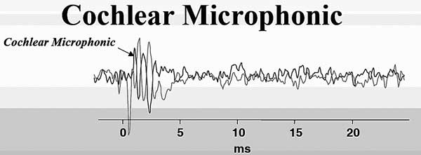
The cochlea generates electrical responses which are most commonly measuring using electrocochleography (in the recent years we haven-t made this test in aur clinic). One of the cochlear potential is the cochlear microphonic CM (Tzenev, 1999), occurring post prior to ABR.

|
|
FIG. 2. Cochlear microphonic |
Cochlear microphonic - CM
not a sign of abnormality, rather an expected response generated in functioning hair cells (both inner and outer).
If seen in an otherwise normal ABR, the CM is not a sign of AN;
CM is due to lock off efferent suppression, not obliterate by the action potential of the auditory nerve, (Starr et all. 2001);
should still be present after OAE disappears and may be the only remaining sign of cochlear function;
ABR changes latency with stimulus level, CM does not;
CM is visible only at high stimulus levels (+ 60 dB).
CM is small in patients with ABR present and larger in patients without ABR;
CM do not change in latency with masking present.
Etiology of Auditory neuropathy:
hereditary, metabolic, toxic of inflammatory neuropothy;
genetics factors Friedreich` s ataxia , Chareot Marie tooth disease, mitochondrial disease;
immune disorders Morbus Grillain Burre;
Infection disease-mumps;
metabolic disorders- hiberbilirubinemia et etc.
Recommended test protocol:
pure tone audiogram;
tympanometry and acoustic reflex (ipsi ad contra);
OAE before ABR;
ABR.
Menagement of Auditory neuropathy:
Hearing aids not a good results, the poor language perception, discrimination particularly in noise;
Cohlear implants good results (similar to the other patients);
- inner hair cell, neurotransmitter, synaptis, losses could leave neural function intact;
- if neural function is affected, then electrical stimulation may still synchronize remaining neural units better than acoustic stimuli.
FM-systems very useful, they help in reducing the back ground noise and minimize the sound to noise ratio.
Communication methods.
Conclusion:
1. Problems in differentiation of neural vs. sensory deafness during screening;
2. The role of OAE in the hearing impaired (lee 2001)
3. The diagnosis of AN should not be a immediate referral for CI;
Improved or fluctuating auditory responses with increasing ages is possible.
REFERENCES
Tzenev, I, Clinic Morphological Otology, Sofia, 1999
Wood S, Mason S, Farnsworth A, et al. Anomalous screening outcomes from click-evoked otoacoustic emissions and auditory brainstem response tests. Br J Audiol 1998;32:399410.
Sininger YS, Starr A. Auditory neuropathy: a new perspective on hearing disorders. San Diego, CA: Singular Publishing, 2001.
Rance G, Beer DE, Cone-Wesson B, et al. Clinical findings for a group of infants and young children with auditory neuropathy. Ear Hear 1999;20:23852.
Starr A, Picton T, Sininger Y, et al. Auditory neuropathy. Brain 1996;119:74153.
Madden C, Hilbert L, Rutter M, et al. Pediatric cochlear implantation in auditory neuropathy. Otology and Neurotology 2002;23:1638.
Shallop JK, Peterson A, Facer GW, et al. Cochlear implants in five cases of auditory neuropathy: postoperative findings and progress. Laryngoscope 2001;111(4 pt 1):55562.
Berlin CI. Auditory neuropathy, using OAEs and ABRs from screening to management. Seminars in Hearing 1999;20:30814.
Davis A, Bamford J, Wilson I, et al. A critical review of the role of neonatal hearing screening in the detection of congenital hearing impairment. Health Technol Assess (Southampton, UK) 1997;1:iiv,1176.
|
Pro Otology |
Journal Home Contents Preview Next |