
|
Journal Home Contents Preview Next |
Pro Otology
Balkan Journal of Otology & Neuro-Otology, Vol. 4, No 2-3:93—95 © 2004
All rights reserved. Published by Pro Otology Association
Modern Concepts in Myringoplasty
V. Sitnikov, B. Zavarzin, I. Chernushevich, V. Kuzovkov
Saint-Petersburg Research Institute of ENT and Speech Disorders, St. Petersburg, Russia
ABSTRACT
Objective: The goal of our study was to compare morphological and functional efficacy of three surgical techniques of myringoplasty.
Study design: The study design is a retrospective review. All techniques are described step by step.
Settings: St.-Petersburg Research Institute of ENT and Speech Disorders, Saint-Petersburg, Russia.
Patients: The study included 123 myringoplasties on 117 patients with uni- or bilateral tympanic membrane perforations.
Interventions: Each patient was operated on according to one of three techniques of myringoplasty. Postoperative examination was performed in 2 weeks, one month and 6 months after operation.
Main outcome measures: Postoperative otolaryngological (including oto-microscopy) examination, pure tone audiometry, tympanometry.
Results: All techniques of myringoplasty we used showed good morphological results. The best results were obtained in cases with two-layer graft closure of subtotal and total timpanic membrane perforations.
Conclusions: Two-layer graft technique might be a good choice for a myringoplasty in cases of different tympanic membrane perforation size. However, medial and “wave-like” techniques give satisfactory results especially in cases with small or medium-sized defects of tympanic membrane.
Key words: Myringoplasty, Surgical technique, Perforation, Tympanic membrane, Two-layer graft.
Pro Otology 2-3:93—95, 2004
INTRODUCTION
From the 50s-60s the otosurgeons started active use of two-layer grafts for myringoplasty aiming the reconstruction of both the outer and the inner surfaces of neotympanic membrane (3,4). Some authors (5,6) described the use of auto- and allocartilaginous plates as a support for facial graft.
Ultrathin allogeneic cartilage, which benefits have been clearly demonstrated by Sitnikov and Kin (1985, 1990) (7), is used for myringoplasty in the past 10-15-years. The benefits are as follows:
1. Semitransparent ultrathin allocartilage allows the surgeon to see the grafts’ bed and therefore to control the correct positioning of the graft.
2. Perforated surface of the ultrathin cartilage favours the diffusion of the nutrients, which is essential for the trophism of the fascial flap.
3. Cartilaginous plate imitates the elastic layer of the tympanic membrane, which accounts for the oscillation capability of the membrane; the plate preserves the anterior meatotympanal angle; it prevents the displacement of the graft towards the tympanic cavity, cartilaginous plate is easy to shape during the insertion.
It is believed that the best results of myringoplasty both from morphological and functional points of view could be achieved in patients with size of the tympanic membrane perforations from small to moderate. The percentage of the complications is significantly higher in patients with subtotal tympanic membrane defects. The most common complications are necrosis, graft rejection, incomplete perforation repairment, and medial graft displacement (2).
PATIENTS AND METHODS
“Underlay” technique – the transplant is placed underneath the medial surface of the tympanic membrane or its remnant.
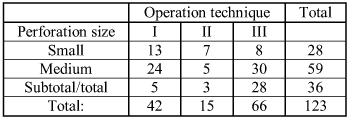
| Table 1. The distribution of the patients according to the size of the perforation. |

|
“Wave-like” technique - transplant is placed in such a way that the anterior portion of the graft is fixed to the medial surface of the tympanic memrane while the posterior portion is placed upon the external surface of the tympanic membrane remnant.
“Onlay two-layer” technique – ultrathin allocrtilaginous plate, which imitates fibro-elastic layer of the destroyed part of the tympanic membrane, and temporal muscle fascia are used.
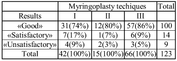
| Table 2. The morphological results |

|
The 3rd operative technique included several stages. In the begnning the epithelium was removed from the remnant of the tympanic membrane. Then so-called ‘wound bed’ was formed, onto which the ultrathin cartilaginous allotransplant was placed. The size of the cartilage was large enough to cover the entire perforation of the tympanic membrane. Upon the cartilage the fascial flap (which was obtained beforehand from the temporal muscle and slightly dryed in order to facilitate the insertion) that exceeded the cartilage in diameter by approximately 1-2mm was placed. Autologous temporal fascial flap was applied then the other two operative techniques were used as well. The fixation of the graft according to the 1st and 2nd operation techniques was performed using a piece of pergameneous paper, which was placed above the fascial flap. A silk thread curled into a spiral was placed above the paper. According to the 3rd techique the fixation of the graft was made by means of ovoid perforated (to enable liquid flow from the wound) latex protector. External auditory meatus was filled with pieces of hemostatic sponge, moistened with saline and antibiotics. By completion of the procedure wound was dressed with a common aseptic bandage. The pieces of the hemostatic sponge were removed from the external auditory meatus in 8-10 days. Patients were enrolled in otu-patient follow-up. The first otomicroscopy of the neotympanic membrane was carried out within 10 days after the operation, the second one – within 15 days, and the next one – within a month. The third examination was usually accompanied by control audiometry. The final assessment of the results of myringoplasty was done in 5-6 months after the operation.
The size of the perforation was the key factor in determining the use of the certain operative technique. The patients were distributed into three groups according to the defect size:
Medium sized (59 cases)
Subtotal and total perforation (36 cases)
The distribution of the patients according to the size of the perforation is shown in Table 1.
As you can see from the table above, the underlay myringoplasty technique (1st) was the most commonly used one (46% of the cases) in patients with small perforation. In this group of patients ‘wave-like’ (2nd) and onlay two-layered (3rd) techniques were used at similar frequency (25% and 29% of the cases respectively), which is almost 2 times less than for 1st techique. As for medium and subtotal/total sized perforations, 3rd (51%) and 1st (41%) techiques were used in the majority of the cases. The share of ‘wave-like’ technique was only 8% in this group of patients. In patients with subtotal perforation the surgeons most frequently used two-layered transplant (3rd). 78% of the patients in this group were operated on using this techique. The shares of 1st and 2nd techniques in this group of patients were 14% and 8% of the cases respectively.
| Table 3. The analysis of the results of the different myringoplasty techniques. |

|
RESULTS
Complete incomplete closure of the perforation.
Elasticity of the neotympanic membrane.
The closure of the perforation was assessed during otoscopy including the evaluation under operation microscope. The elasticity of the neotympatic membrane was assessed with tympanometry.
To grade the morphological results we used the following definitions:
«Good» - Complete closure of the perforation without any signs of displacement of the flap or adhesion of the flap to the medial wall of the tympanic cavity.
«Satisfactory» - The area of the perforation has decreased by 50% or more or there is a medial displacement of the flap while the perforation has closed completely.
«Unsatisfactory» - The signs of the total or partial graft rejection are observed.
The morphological results are presented in Table 2.
As you can see from the table above, even if we don’t take into account the size of the perforation, we have to conclude that the 3rd myringoplasty technique (which uses two-layer transplant) is the most effective one. On the other hand, the other two techniques provide high percentage of positive results (74-80%).
Table 3 presents the analysis of the results of the different myringoplasty techniques, which takes into account the size of the perforation of the tympanic membrane. Two-layer graft technique has shown to produce high percentage of positive results in all groups of patients: 100% in patients with small perforations, 90% in patients with medium-sized perforations, and 78% in patients with total/subtotal perforations. Unsatisfactory result of this technique was observed in the following distinct instances: 1 patient with medium-sized perforation and 2 patients with subtotal perforation.
Tympanometry was performed only in patients with good morphological results. According to the obtained data the best results have been registered in patients operated on using the 3rd myringoplasty technique.
CONCLUSIONS
REFERENCES
Barteneva AA, Kozlov MY. Problems of tympanoplasticy. L. 1974:84-6.
Tarasov DI. Disease of the middle ear. M., Medicine , 1988
Wullstein H. Theory and practice of tympanoplasty. Laryngoscope 1956;66:1076-95.
Mishenkin NV, Sintikov VP. New kind of transplantation of ossiculs by patiens with chronical otitis media. Omsk, 1975:13-6.
Austin DF. Reporting results in tympanoplasty. Am J Otol 1985;6:85-8.
Qshan IA, Rishko NM. Endomeatal tympanoplasticy with fixation of plastic metal prothesis. 2-nd otorhinolaringological congress Byelorussia, Minsk, High school, 1984:89-90.
Sitnikov VP, Kin TI. Myringoplastic by patients with big defect of the tympanic membrane. Method recommendation. M., 1990:22-5.
|
Pro Otology |
Journal Home Contents Preview Next |