|
|
Journal Home Contents Preview Next |
Pro Otology
Balkan Journal of Otology & Neuro-Otology, Vol. 4, No 2-3:8792 © 2004
All rights reserved. Published by Pro Otology Association
Otogenic Sinus Thrombosis and Hydrocephalus
after Acute Otitis Media in Pediatric Patients
*A. Koitschev, *H. Loewenheim, *Chr. Simon, *M. Kumpf, D. Besch, U. Ernemann
*Department of Otorhinolaryngology and Head and Neck Surgery, University of Tuebingen, Germany
Department of Ophthalmology, University of Tuebingen, Germany
*Department of Pediatrics II, University of Tuebingen, Hoppe-Seyler-Str. 3, D-72076, Germany
Department of Neuroradiology, University of Tuebingen, Hoppe-Seyler-Str. 3, D-72076, Germany
ABSTRACT
Objectives: To describe the frequency, pathognomonic signs, clinical course and outcome of otogenic dural sinus thrombosis (DST) as an intracranial complication of an acute otitis media (AOM) in pediatric patients.
Study Design, Setting, and Patients: Retrospective cohort review of all pediatric patients (age 1-14 years) between1999 and 2004 treated for otitis media and complications of it in an academic medical center. Otologic and ophthalmologic findings and CT and MRI imaging at the beginning of treatment and 3 months later were analyzed.
Results: We report on 6 cases with otogenic DST following AOM. Three of them were female and three male. The age was between 3 and 14 years. Four of them primarily presented with diplopia. In these cases the otologic complaints had already disappeared at the time of MRI confirmation of the diagnosis. Two children were primarily referred with otologic symptoms.
The management included systemic antibiotics, short time heparin anticoagulation, and surgical decompression. In our cases, even after intensive i.v. antibiotics, only surgical treatment lead to significant improvement of the clinical condition.
Conclusions: The clinical presentation of otogenic DST as a complication from AOM could be masked due to antibiotic treatment. Morning episodes of vomiting and/or headache, visual impairment, and a history of AOM seem to be indicative for otogenic hydrocephalus in cases without otologic symptoms. We advocate therefore a generous consideration of MRI in patients with similar symptoms to facilitate an early diagnosis of a potentially lifethreatening complication.
Key words: Pediatric, Otogenic sinus thrombosis, Acute otitis media, MRI.
Pro Otology 2-3:8792, 2004
Introduction
Acute otitis media (AOM) is one of the most common diagnoses in children. Despite controversial discussion of antibiotic treatment of uncomplicated AOM (1), compared to the pre-antibiotic era (2) severe complications of AOM such as osteomyelitis, meningitis, brain abscess and dural sinus thrombosis (DST) have become very rare today. If they occur, antibiotic coverage may not have been sufficient. Typical uncomplicated pediatric AOM with otalgia, hearing impairment and severe illness is an acute condition, which is, despite some controversies in the definition of the disease (1), easily recognized by an experienced clinician.
However the clinical presentation of complications from AOM could be masked due to the antibiotic treatment. We report on 6 children with otogenic DST. 4 of them presented with bilateral papilloedema due to increased intracranial pressure causing diplopia. All of them had a history of clinically successfully treated AOM 1 to 4 weeks prior to the onset of visual symptoms. In 4 of the 6 cases the otologic complaints had already disappeared at the time of MRI confirmation of the diagnosis.Methods
Charts of pediatric patients (age 1-14 years) between1999 and 2004 treated for otitis media and complications of it were retrospectively reviewed. Only patients with sinus thrombosis confirmed by CT or MRI and ipsilateral mastoiditis were included in the series. Otological and ophthalmological findings, operative reports, radiological examinations, laboratory data, medical treatment and outcome were investigated
Results
6 pediatric patients with otogenic DST were identified. Three of them were female and three male. The age was between 3 and 14 years.
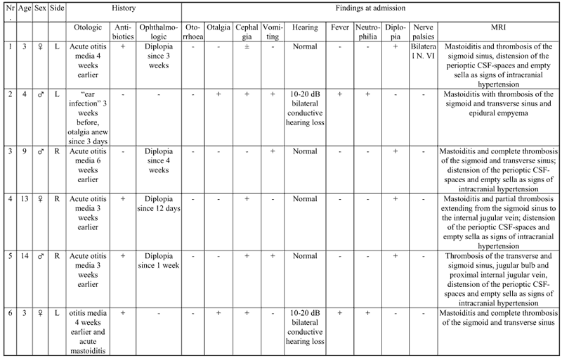
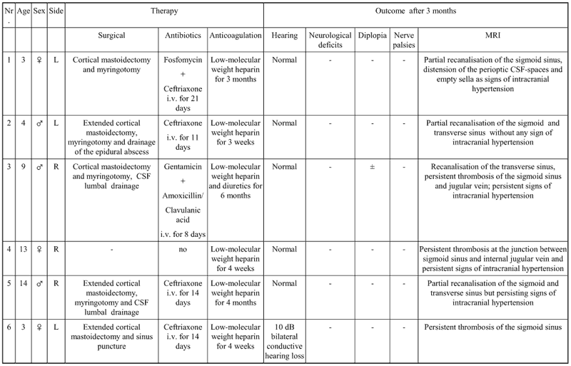
Magnetic resonance imaging (MRI) revealed in all cases unilateral mastoiditis and thrombosis of at least one neighboring dural sinus. Four of the patients were initially seen by an ophthalmologist due to diplopia. Two children were referred with severe otologic symptoms. These cases did not present ophthalmologic impairment. Blood cultures (case 2) prior to the initiation of systemic antibiotic therapy revealed an infection with Haemophilus influenca. In all but one (case 6) patients intraoperative cultures from the mastoid cavity were negative for bacterial infections. In case number 6 microbiology has shown Streptococcus pneumoniae and Neisseria species. Tables 1 and 2 summarize the medical history, findings on admission, therapy and outcome of all patients. The following case reports were chosen as a representative description of the clinical presentation of otogenic DST with (case1) and without (case 2) hydrocephalus and ophthalmologic symptoms.| Table 1. History and findings at admission. |
|
Case reports
Otogenic DST with hydrocephalus (case 1)
A 3-year-old girl was referred with diplopia caused by bilateral papilloedema with reduced visual acuity on the left side and beginning bilateral sixth cranial nerve palsies. She had presented four weeks previously with left otalgia, headache and fever up to 40° C and was diagnosed with a left sided AOM by her general practitioner. Oral treatment with penicillin had been clinically successful. She had had no previous history of otological problems. During the weeks after the AOM a progressive left eye winking and head tilting occurred, which led to an ophthalmologic referral. On the day of admission neither fever nor neutrophilia were present. The left tympanic membrane was slightly erythematous and retracted with minimal effusion.
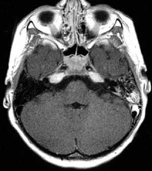
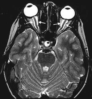
Computed tomography (CT) showed a total opacification of the left mastoid as a sign of mastoiditis. MRI revealed a partially recanalised thrombosis of the left sigmoid sinus (FIG. 1A) as well as prominent perioptic cerebro-spinal fluid (CSF) spaces and empty sella indicating intracranial hypertension (FIG. 1B). There were no signs of meningitis or cerebral inflammation. Antibiotics (fosfomycin and ceftriaxone) and low-molecular weight heparin (LMWH) anticoagulation treatment were started. After a week of conservative therapy without improvement of the papilloedema a cortical mastoidectomy and myringotomy on the left side were performed. Seromucotympanon and mastoiditis with granulations but no abscess formation was found. The postoperative recovery was unremarkable with continuous improvement of diplopia. Four months later diplopia and otologic symptoms had completely resolved, but MRI signs of intracranial hypertension and reduced visual acuity on the left side persisted.Otogenic DST without hydrocephalus (case 2)
A 4-year-old boy was referred with a three day history of left sided otalgia, pyrexia up to 40° C and vomiting. He had been prescribed ear drops for ipsilateral ear infection three weeks earlier.
On examination he had fever spikes up to 40° C without clinical mastoiditis signs. The left tympanic membrane was red and thickened. Under the diagnosis of left AOM he was started on intravenous cefotiam. Since fever and neutrophilia persisted despite the antibiotic treatment imaging studies were performed. CT and MRI revealed the intracranial complication of mastoiditis with thrombosis of the sigmoid and transverse sinus and an epidural empyema. Haemophilus influencae was isolated in the blood culture. Extended cortical mastoidectomy, myringotomy and drainage of the epidural abscess were performed and change of antibiotic to ceftriaxone was made. Over the next week the clinical picture returned completely to normal. Three months later an MRI showed partial recanalisation of the sigmoid and transverse sinus without any sign of intracranial hypertension.| Table 2. Therapy and outcome. |
|
Discussion
Widespread use of antibiotics in pediatric cases of AOM has considerably diminished the incidence of intracranial complications such as DST, which was common in the pre-antibiotic era (2). Earlier publications reported series of such cases with a mortality rate of up to 90 % (2-4). Many of those cases were complicated by a chronic otitis media with cholesteatoma and are therefore not comparable to a common AOM (5). Today this potentially lethal condition still occurs(6,7), although in a changed clinical appearance.
Otogenic DST results from direct spread of infection from the mastoid and used to be a severe illness with septic fever and signs of meningitis. This classical clinical picture, including otologic symptoms was present in two cases of our series. In contrast to most of the previous reports of otogenic DST, the majority of our patients did not present with acute otologic symptoms at the time of diagnosis. The main symptom was diplopia and edema of the optic discs as a sign of increased intracranial pressure, combined with episodes of headache and vomiting. The patients had no other otologic medical history other than being successfully treated for a AOM a few weeks prior to presentation. None of them had chronic otitis media. Routine laboratory evaluation for prothrombotic disorders was negative. Contrast enhanced CT and MRI phlebography established the diagnosis and were shown to be sensitive in staging intracranial complication of AOM. CT and MRI were successfully performed under sedation in all cases. The management of otogenic DST includes antibiotics, short time heparin anticoagulation and surgical decompression. In our cases, even after intensive i.v. antibiotics, only surgical treatment lead to significant improvement of the clinical condition. This is in contrast to a recently published series consisting of four children with DST, whose clinical symptoms resolved with conservative management only. Of note, the children in this series had no visual impairments suggesting a lesser degree of severity of their intracranial complication (9). In only one of the patients cultures from the mastoid cavity returned bacterial growth. DSTs therefore developed (except case 6) despite sufficient elimination of the specific infection through systemic antibiotic therapy. This circumstance needs special attention, because it requires the physician to suspect severe intracranial complications of AOM without otologic symptoms. The role of anticoagulation remains unclear. Even with low-molecular weight heparin anticoagulation performed, only partial recanalisation of the affected sinuses and persisting signs of intracranial hypertension were observed by MRI three months after treatment. These cases demonstrate that normal otoscopic findings do not exclude a severe intracranial complication of AOM and highlight the distinctly altered and delayed clinical appearance of DST as a complication of AOM after antibiotic treatment. During mastoidectomy the surface of the sigmoid sinus was explored. Only one case (case 6) presented signs of thrombophlebitis or osseous erosion. Therefore puncture of the sigmoid sinus was performed only in that case. We may speculate that an aseptic mastoiditis has lead to a mural DST with consecutive hydrocephalus in cases of visual impairment. This condition is known as otitic hydrocephalus (10,11)being a subcategory of larger entity named pseudotumor cerebri (12) . The pathophysiology of otitic hydrocephalus remains controversial. It has been argued that involvement of the superior sagittal sinus is a necessary component of this disease. In our cases of otitic hydrocephalus the MRI revealed normal luminal and mural flow within the superior sagittal sinus. The presence of thrombus in the lateral venous sinus alone appears sufficient to produce a rise in the cerebral venous pressure and a subsequent increase in the CSF pressure. Imaging such as high resolution MRI may give new insights into this rare complication of otitis and an opportunity to differentiate from similar conditions like pseudotumor cerebri. Morning episodes of vomiting and/or headache, visual impairment, and a history of AOM seem to be indicative for otogenic DST with hydrocephalus. We advocate therefore a generous consideration of MRI in patients with similar symptoms even without otologic symptoms to facilitate an early diagnosis of a potentially lifethreatening complication.REFERENCES
Takata GS, Chan LS, Shekelle P, Morton SC, Mason W, Marcy SM. Evidence assessment of management of acute otitis media: I. The role of antibiotics in treatment of uncomplicated acute otitis media. Pediatrics. 2001;108:239-47.
Newton Pitt, G. An analysis of fifty-seven fatal cases of ear disease, and of the complications which led to death. BMJ 1, 1980;22(3):643-7.
Nielsen, JM. and Courville, CB. Intracranial complications of otogenous thrombosis of the lateral sinus. Ann. Otol. Rhinol. Laryngol. 1937;46:12-38.
Sutherland, J. M. Otitic sinus thrombosis. Arch. Otolaryngol. 1938;27:1-34.
Kaplan DM, Kraus M, Puterman M, Niv A, Leiberman A, Fliss DM. Otogenic lateral sinus thrombosis in children. Int J Pediatr Otorhinolaryngol. 1999;49:177-83.
Jose J, Coatesworth AP, Anthony R, Reilly PG. Life threatening complications after partially treated mastoiditis. BMJ. 2003; 327:41-2.
Garcia RD, Baker AS, Cunningham MJ, Weber AL. Lateral sinus thrombosis associated with otitis media and mastoiditis in children. Pediatr Infect Dis J. 1995;14:617-23.
Dobben GD, Raofi B, Mafee MF, Kamel A, Mercurio S. Otogenic intracranial inflammations: role of magnetic resonance imaging. Top Magn Reson Imaging. 2000;11:76-86.
Rocha JL, Kondo W, Gracia CM, Baptista MI, Buchele G, da Cunha CA et al. Central venous sinus thrombosis following mastoiditis: report of 4 cases and literature review. Braz J Infect Dis. 2000;4:307-12.
Symonds, C. P. Otitic hydrocephalus. BMJ 1932;1:53-5.
Tomkinson A, Mills RG, Cantrell PJ. The pathophysiology of otitic hydrocephalus. J Laryngol Otol. 1997;111:757-9.
Johnston IH, Duff J, Jacobson EE, Fagan E. Asymptomatic intracranial hypertension in disorders of CSF circulation in childhood-treated and untreated. Pediatr Neurosurg. 2001;34:63-72.
|
Pro Otology |
Journal Home Contents Preview Next |